
Understanding the precise chemical makeup of historical pigments offers deep insights into artistic practices, materials science, and even trade routes of the past. This knowledge helps conservators make informed decisions about preserving priceless artworks and artifacts. Energy-dispersive X-ray spectroscopy elemental pigment mapping conservation provides a non-destructive or minimally destructive way to achieve this detailed understanding.
This powerful analytical technique allows researchers to identify the elements present in a pigment and map their distribution across a sample. The resulting elemental maps reveal how artists layered colors or mixed different materials, offering clues about their creative process. EDX pigment analysis has become an indispensable tool for cultural heritage scientists.
By revealing the elemental composition, we can confidently identify specific pigments, differentiate between original materials and later restorations, and even detect forgeries. The ability to perform elemental mapping paintings with high spatial resolution transforms our understanding of artistic masterpieces. It truly is a game-changer for SEM-EDX art conservation.
How EDX Works and Why It Is Paired With Scanning Electron Microscopy
Energy-dispersive X-ray spectroscopy, or EDX, operates on a fundamental principle of atomic physics: the interaction of electrons with matter. When a high-energy electron beam strikes a sample, it excites atoms within the material. This excitation causes inner-shell electrons to be ejected from their orbits.
To stabilize, an electron from a higher energy shell fills the vacant inner-shell position. This transition releases energy in the form of a characteristic X-ray photon. Each element emits X-rays with unique energy levels, acting like a fingerprint.
An EDX detector collects these X-ray photons and sorts them by their energy. This creates a spectrum where peaks correspond to specific elements present in the sample. The intensity of each peak relates to the concentration of that element, giving both qualitative and quantitative information.
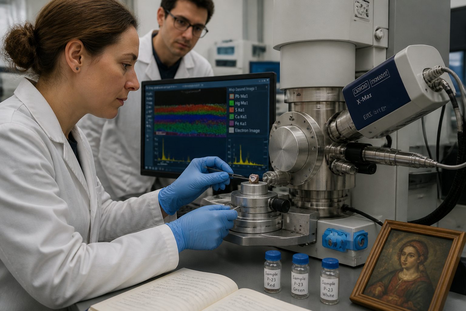
EDX is almost always paired with a scanning electron microscope (SEM) because the SEM provides the focused electron beam necessary for excitation. The SEM also generates high-resolution images of the sample surface, allowing researchers to correlate elemental data with specific morphological features. This combination, often called SEM-EDX art conservation, is incredibly effective.
The finely focused beam of the SEM allows for precise targeting of microscopic pigment particles or layers within a cross-section. This spatial resolution is critical for understanding complex stratigraphies in paintings. It ensures that the energy dispersive X-ray spectroscopy elemental pigment mapping conservation is accurate and specific to the areas of interest.
What Elements EDX Can Detect and What That Tells You About Pigments
EDX pigment analysis can detect elements from beryllium (atomic number 4) up to uranium (atomic number 92), covering most elements relevant to historical pigments. This wide range means we can identify nearly all inorganic compounds used as colorants throughout history. Organic pigments, which lack heavy elements, are generally not detectable by EDX directly.
The presence of specific elements immediately points to certain pigments. For example, detecting lead (Pb) and tin (Sn) together usually indicates lead-tin yellow, a common pigment from the medieval period through the 18th century. Similarly, high levels of copper (Cu) might suggest azurite or malachite, both copper carbonates used for blue and green colors.
Iron (Fe) is a ubiquitous element found in earth pigments like ochres, siennas, and umbers, which vary in color based on their iron oxide and hydroxide forms. The presence of mercury (Hg) and sulfur (S) strongly points to vermilion, a synthetic or naturally occurring red pigment. Knowing these elemental signatures is fundamental to energy dispersive X-ray spectroscopy elemental pigment mapping conservation.
Even trace elements can be informative, sometimes indicating the geological origin of a natural pigment or impurities introduced during manufacture. For instance, the presence of specific minor elements in a lead white sample might help distinguish between different historical production methods. This level of detail helps conservators understand provenance and authenticity.
Beyond individual pigments, elemental mapping paintings can reveal mixtures of pigments. If a green area shows both copper (from a blue pigment) and iron (from a yellow ochre), it suggests the artist mixed a blue and a yellow to achieve the green. This insight into artistic technique is invaluable for art historians and conservators alike.
Sample Preparation Requirements for SEM-EDX Analysis
Proper sample preparation is absolutely critical for obtaining accurate and meaningful results from SEM-EDX analysis. Poor preparation can introduce artifacts, obscure important features, or even damage the sample. Conservators must balance the need for minimal intervention with analytical requirements.
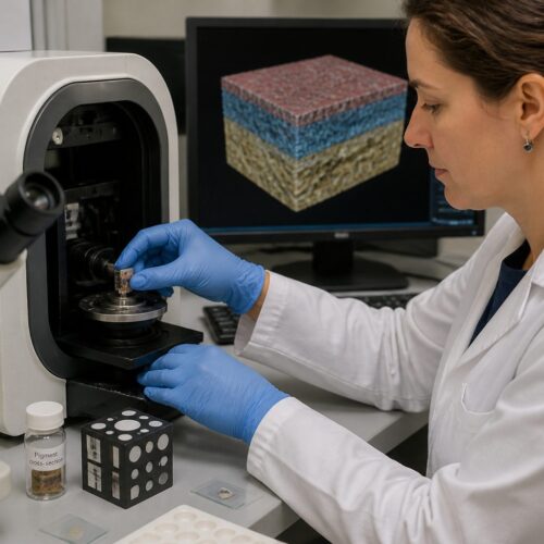
For paintings, the most common sample type is a microscopic cross-section, typically a tiny fragment removed from a non-visible or already damaged area. This fragment is embedded in a resin, then polished to reveal the stratified layers of paint. Preparing these cross-sections demands extreme care and precision.
| Preparation Step | Purpose | Considerations for Pigments |
|---|---|---|
| Sampling | Obtain representative, minimal material | Choose inconspicuous areas, use sterile tools, record location |
| Embedding | Support fragile layers, create a flat surface | Use transparent, low-Z resin; avoid air bubbles |
| Polishing | Expose a smooth, cross-sectional view | Use fine abrasives, avoid smearing soft layers, maintain layer integrity |
| Carbon Coating | Ensure conductivity, prevent charging | Apply thin, uniform layer; avoid heavy element contamination |
| Cleaning | Remove surface contaminants | Use mild solvents or air; prevent material loss |
The sample must be electrically conductive to prevent charging under the electron beam, which can distort images and spectra. Non-conductive samples, like most paint layers, are typically coated with a very thin layer of carbon or a noble metal like gold or platinum. Carbon is preferred for EDX pigment analysis because it introduces no heavy elements that could interfere with pigment detection.
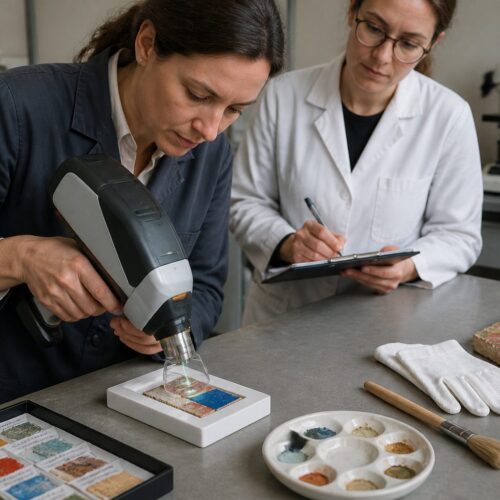
Sometimes, analysis can be performed directly on a surface, such as a microscopic area of a sculpture or ceramic, without removing a sample. This “in situ” approach is less invasive but often provides less detailed stratigraphic information. The choice of preparation method depends on the artifact, the research question, and the ethical constraints of SEM-EDX art conservation.
The size of the sample is usually very small, often less than a millimeter, making the removal almost imperceptible to the naked eye. Despite their tiny size, these samples can hold a wealth of information about the artwork’s history. This meticulous preparation ensures the high quality of elemental mapping paintings.
Reading EDX Spectra and Element Maps for Pigment Identification
Interpreting EDX spectra involves identifying peaks corresponding to characteristic X-ray energies for each element. The software typically labels these peaks, making initial identification straightforward. However, overlapping peaks or background noise can sometimes complicate interpretation, requiring an experienced eye.
Beyond simply identifying elements, the relative heights of the peaks provide an indication of elemental concentrations. Stronger peaks mean more of that element is present. This quantitative aspect helps distinguish between a primary pigment component and a minor impurity.
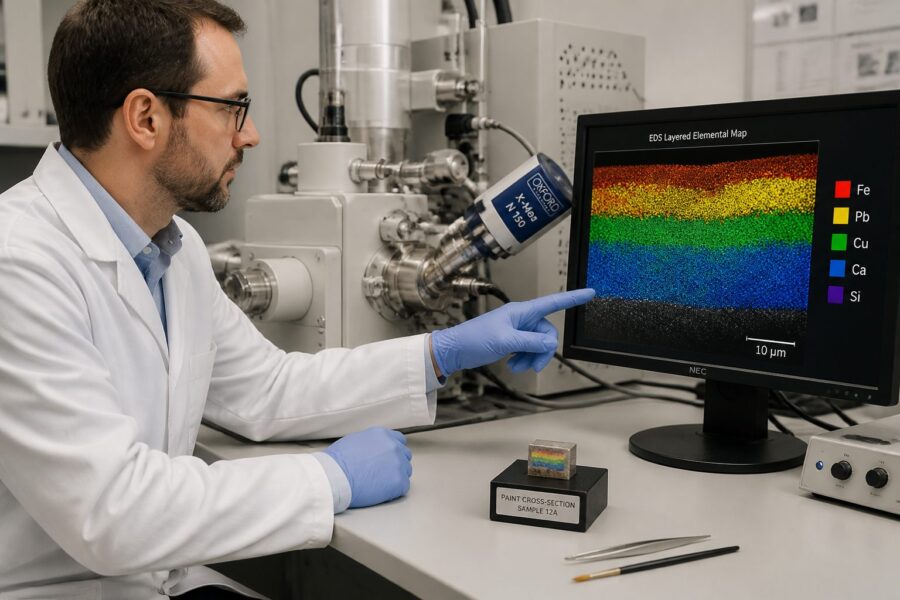
Element maps are arguably the most visually compelling output of energy dispersive X-ray spectroscopy elemental pigment mapping conservation. These maps show the spatial distribution of selected elements across the analyzed area, usually overlaid on the SEM image. Each element is assigned a different color, allowing for easy visualization of elemental segregation and co-localization.
For example, an element map of a painting cross-section might show a distinct layer of lead (Pb) and sulfur (S) in one section, indicating vermilion. Directly above it, a layer showing calcium (Ca) and carbon (C) could point to a chalk ground layer. These maps literally make the invisible layers visible.
Interpreting these maps requires knowledge of historical pigments and their typical elemental compositions. You look for elements that commonly occur together in known pigments. Seeing copper and chlorine together might suggest atacamite, a specific copper chloride pigment, rather than a simple copper carbonate like azurite. This detailed analysis is what makes elemental mapping paintings so powerful for conservation.
Case Studies Where EDX Resolved Ambiguous Pigment Identifications
EDX pigment analysis has repeatedly proven its worth by clearing up ambiguities in art historical and conservation studies. Its ability to provide elemental specifics often settles debates about material authenticity or artistic technique. These real-world applications demonstrate the power of energy dispersive X-ray spectroscopy elemental pigment mapping conservation.
One notable case involved distinguishing between natural ultramarine and Prussian blue, two blue pigments with similar visual appearances but vastly different compositions and historical periods. Natural ultramarine is a sulfur-containing sodium-aluminum silicate, while Prussian blue is an iron ferrocyanide. EDX easily differentiates them by detecting sulfur, sodium, and aluminum in the former and iron in the latter, without any of the other elements.
- Confirming authenticity of ancient Egyptian blue
- Identifying cochineal lake pigments via aluminum and potassium
- Differentiating between lead-tin yellow Type I and Type II
- Detecting early use of cadmium yellow in 19th-century works
- Revealing hidden layers of original pigments beneath restorations
- Pinpointing specific iron oxides in different ochre varieties
- Distinguishing between natural cinnabar and synthetic vermilion
Another common ambiguity EDX resolves involves white pigments. Lead white (lead carbonate) was widely used for centuries, but zinc white (zinc oxide) and titanium white (titanium dioxide) became popular later. EDX clearly identifies lead, zinc, or titanium, allowing conservators to date paint layers or detect anachronistic materials. This helps in authenticating artworks and understanding their material history.
In a study of a Renaissance painting, SEM-EDX art conservation revealed that a vibrant green color, previously assumed to be malachite, actually contained copper and arsenic, identifying it as Scheele’s green or Paris green. This discovery changed the understanding of the artist’s palette and the potential toxicity risks during conservation. Elemental mapping paintings provide undeniable proof.
EDX also helped identify a rare pigment, orpiment (arsenic trisulfide), in a medieval manuscript where its presence was unexpected. The detection of arsenic and sulfur in the yellow areas provided definitive proof, offering new insights into the manuscript’s production. These examples show how EDX helps redefine our understanding of art history.
These case studies underscore the method’s power in art conservation. They confirm that EDX is not just a theoretical tool, but a practical one that delivers concrete answers. It continues to be a cornerstone for material analysis in museums and conservation labs worldwide.
Comparing EDX With XRF: Spatial Resolution and Depth Sensitivity
When discussing elemental analysis in art conservation, X-ray fluorescence (XRF) is another widely used technique, and it’s important to understand how it compares to EDX. Both methods provide elemental information, but they differ significantly in their operational principles, spatial resolution, and depth sensitivity. This comparison helps clarify when to choose one over the other for energy dispersive X-ray spectroscopy elemental pigment mapping conservation.
XRF uses X-rays to excite the sample, causing it to emit secondary X-rays that are characteristic of its elemental composition. Unlike EDX, XRF does not require a vacuum environment, making it a truly non-contact and often portable technique. This portability is a huge advantage for analyzing large, immovable objects directly on site.
The primary difference in spatial resolution is stark. Laboratory-based XRF can analyze areas down to a few hundred micrometers, while portable XRF typically has a spot size of several millimeters. In contrast, SEM-EDX art conservation can achieve spatial resolution down to the nanometer scale, thanks to the finely focused electron beam of the scanning electron microscope. This means EDX can probe individual pigment particles or extremely thin paint layers with precision.
Regarding depth sensitivity, both techniques are surface-sensitive but differ in their penetration depth. XRF typically penetrates deeper into the sample, analyzing a volume ranging from tens to hundreds of micrometers, depending on the element and matrix. This means XRF provides an average elemental composition of multiple paint layers.
EDX, on the other hand, typically analyzes a much shallower depth, usually a few micrometers, due to the limited penetration of the electron beam. This shallower analysis depth can be an advantage when studying very thin surface layers or individual particles. However, it also means EDX might not detect elements from deeply buried layers without preparing a cross-section. Elemental mapping paintings with EDX offers more precise layer-by-layer details.
Therefore, XRF is excellent for rapid, non-invasive screening of large areas and for identifying major elements in bulk materials. EDX pigment analysis is superior for detailed, high-resolution investigation of micro-samples, especially when precise spatial distribution and stratigraphic information are needed. Often, conservators use XRF for initial surveys and then follow up with EDX on micro-samples for targeted, detailed analysis.
Limitations and Where EDX Falls Short as a Standalone Method
While energy dispersive X-ray spectroscopy elemental pigment mapping conservation is incredibly powerful, it does have limitations that prevent it from being a standalone method for comprehensive material characterization. Understanding these shortcomings helps integrate EDX effectively with other analytical techniques. No single method provides all the answers.
One major limitation is its inability to detect very light elements, specifically hydrogen, helium, lithium, and sometimes beryllium and boron, with good sensitivity. This means EDX cannot directly analyze the organic binders, varnishes, or most organic pigments that constitute a significant part of historical artworks. These organic components are crucial for understanding the overall composition and degradation processes.
Another drawback is its destructive or minimally destructive nature, especially when micro-samples are required. While the samples are tiny, removing material from an artwork is always a significant decision and not always permissible for highly sensitive or intact objects. This contrasts with truly non-invasive techniques like portable XRF or Raman spectroscopy.
EDX also struggles with distinguishing between different chemical states or crystalline forms of the same element. For instance, it can detect iron, but it cannot tell you if that iron is present as hematite, goethite, or a modern iron oxide pigment. These different forms have distinct optical properties and historical uses, which are important for accurate pigment identification. This is a key area where EDX pigment analysis needs complementary methods.
The quantification of elemental concentrations can also be challenging, especially in complex matrices like paint layers with varying densities and surface roughness. While EDX provides relative concentrations, obtaining highly accurate absolute quantitative data often requires careful calibration and matrix correction. The accuracy can be affected by sample geometry and surface condition.
Finally, the SEM-EDX art conservation process requires a vacuum environment, which means larger artifacts cannot be analyzed directly. Samples must be small enough to fit inside the microscope chamber and withstand the vacuum. This physical constraint dictates the type and size of objects that can be studied. For these reasons, elemental mapping paintings often benefits from a multi-analytical approach.
Conclusion
Energy-dispersive X-ray spectroscopy, particularly when combined with scanning electron microscopy, stands as a cornerstone analytical technique in the field of art conservation. Its ability to provide detailed elemental composition and spatial distribution of pigments has revolutionized how we understand and preserve historical artworks. This powerful method provides unprecedented insights into artistic materials and techniques.
From identifying ancient pigments to detecting modern forgeries, EDX pigment analysis offers invaluable data for conservators, art historians, and scientists. The precise elemental mapping paintings reveal hidden layers, mixtures, and alterations that are invisible to the naked eye. This level of detail ensures informed conservation decisions.
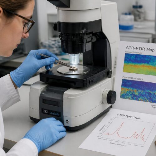
While EDX excels in elemental identification and mapping, its limitations, such as insensitivity to light elements and the need for micro-samples, mean it is best utilized as part of a multi-analytical strategy. Combining EDX with techniques like XRF, Raman spectroscopy, or FTIR provides a more complete picture of an artwork’s material history. This integrated approach maximizes the insights gained from each method.
The ongoing development of EDX technology, including improved detectors and software, continues to enhance its capabilities, making it even more indispensable for cultural heritage science. The future promises even greater resolution and analytical precision. This will further solidify its role in energy dispersive X-ray spectroscopy elemental pigment mapping conservation.
Ultimately, SEM-EDX art conservation allows us to delve deeper into the material stories embedded within our cultural heritage. It helps us reconstruct artistic processes and safeguard these treasures for future generations. The information gained from EDX analysis is a vital part of preserving human creativity.