
Understanding the layers and composition of historical artworks is essential for effective conservation. Conservators and art historians often rely on non-destructive methods to study these precious objects without causing any damage. Multispectral imaging pigment mapping non-invasive techniques offer a powerful solution for this delicate work.
This advanced method allows researchers to see beyond the visible surface, revealing hidden details about pigments and their distribution. It provides critical insights into an artist’s original intent, subsequent alterations, and the condition of the materials. Multispectral pigment analysis has become a cornerstone in the field, guiding difficult conservation decisions with objective data.
The ability to map pigment distribution without even touching the artwork is a game-changer for cultural heritage. This non-destructive art imaging approach preserves the integrity of the object while delivering unprecedented analytical depth. It allows us to ask new questions and find answers that were previously impossible to obtain.
How Multispectral Imaging Works and What It Captures
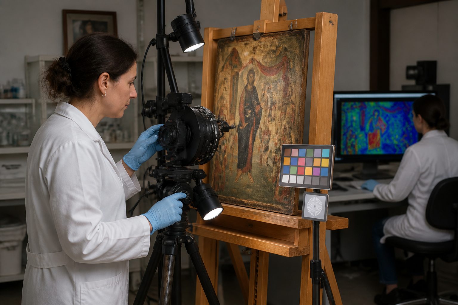
Multispectral imaging involves capturing images of an artwork across many different light wavelengths, far beyond what the human eye can perceive. This process typically uses a specialized camera equipped with filters or a tunable lens to isolate specific bands of light. Each wavelength interacts differently with the materials on the artwork’s surface and subsurface.
When light hits a pigment, it can be absorbed, reflected, or transmitted in various ways depending on the pigment’s chemical composition. By recording how much light is reflected at each specific wavelength, we build a spectral fingerprint for different materials. This unique signature allows us to distinguish between pigments that might look identical under normal visible light.
The system collects a series of grayscale images, each representing the artwork’s reflectance at a narrow band of the electromagnetic spectrum. These individual images are then processed and combined to create a comprehensive dataset. This dataset is the foundation for detailed multispectral pigment analysis, showing variations in material properties.
The technique can reveal underlying sketches, pentimenti, and even previous restoration attempts that are invisible to the naked eye. It provides a detailed map of where specific pigments are located across the surface of a painting or manuscript. This non-destructive art imaging gives conservators a clearer picture of the artwork’s history and construction.
Multispectral imaging pigment mapping non-invasive methods are particularly effective for identifying organic dyes and certain inorganic pigments. It works by exploiting the distinct absorption and reflection characteristics of each material. This allows for the differentiation of pigments that might appear similar in visible light but behave very differently in infrared or ultraviolet regions.
Wavelength Ranges Used and What Each Reveals About Pigments
Different parts of the electromagnetic spectrum offer unique perspectives on an artwork’s composition. Visible light, ranging from about 400 to 700 nanometers, shows us the colors we normally see and gives a baseline for comparison. This range helps us understand how the artwork appears to a typical viewer.
Ultraviolet (UV) light, below 400 nm, often causes certain materials to fluoresce, emitting visible light. This UV fluorescence helps identify organic binders, varnishes, and some pigments, as well as indicating areas of restoration or overpaint. It shows us where different surface coatings or materials might be present.
Infrared (IR) radiation, typically above 700 nm, penetrates deeper into paint layers than visible light. This allows us to see underdrawings, changes in composition, and specific pigments like carbon black, which become transparent in this range. Infrared false-color images are crucial for pigment distribution mapping and revealing hidden elements.
Shortwave infrared (SWIR) extends this penetration even further, offering insights into pigments that are opaque in the near-infrared range. This can be particularly useful for distinguishing between different types of iron-gall inks or certain dark pigments. SWIR imaging helps refine our understanding of complex layered structures.
Some advanced systems also capture images in the terahertz range, which can provide even deeper penetration through multiple layers of paint and support. This emerging technology offers potential for studying very thick paint applications or multi-layered textiles. Each wavelength range adds another piece to the puzzle of an artwork’s construction.
Setting Up a Multispectral Imaging Session in a Conservation Lab
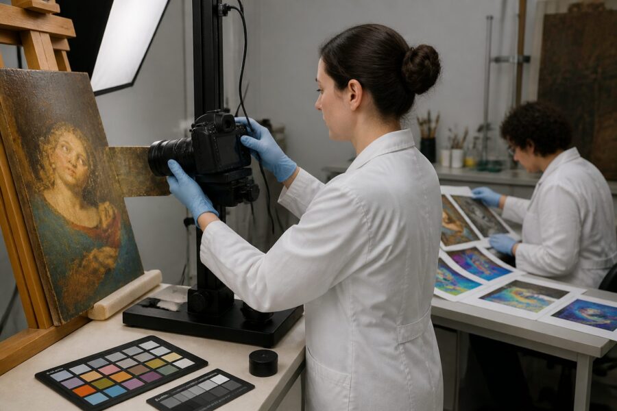
Proper setup is paramount for acquiring high-quality multispectral imaging pigment mapping non-invasive data. The first step involves ensuring a stable environment, controlling both temperature and humidity to protect the artwork. This prevents any environmental stress on the delicate materials during the imaging process.
The artwork needs secure positioning on a stable easel or support, perfectly perpendicular to the camera’s lens. Consistent and even illumination across the entire surface is also critical, usually achieved with multiple balanced light sources. This ensures uniform data capture without shadows or hotspots.
| Equipment Component | Purpose in MSI Setup | Key Considerations |
|---|---|---|
| Specialized Camera | Captures images across specific wavelength bands | High resolution, low noise, spectral sensitivity |
| Tunable Filters/Lenses | Selects precise wavelength ranges for capture | Accuracy of bandwidth, rapid switching capabilities |
| Broadband Light Source | Provides consistent illumination across the spectrum | Uniformity, intensity control, minimal heat emission |
| Calibration Targets | Used for color and reflectance correction | Standardized materials, regular recalibration |
| Computer Workstation | Controls camera, processes, and stores data | High processing power, ample storage, specialized software |
| Art Object Support | Secures artwork in a stable, flat position | Adjustability, non-damaging contact points, vibration dampening |
Interpreting False-Color Maps to Identify Pigment Zones
False-color images are a primary output of multispectral pigment analysis and require careful interpretation. These images assign visible colors to specific wavelength bands or mathematical combinations of them, making invisible spectral differences apparent to the human eye. They allow us to visualize data that would otherwise remain hidden.
For example, an infrared false-color image might assign red to a high reflectance in the IR range, green to visible red, and blue to visible green. This combination can make certain pigments, like Egyptian blue or ultramarine, appear in striking, non-natural colors. These color shifts are not arbitrary; they are based on how specific pigments interact with light.
By comparing the false-color maps with known spectral libraries of historical pigments, conservators can identify specific pigment zones. If an area glows bright blue in an IR false-color image, and that spectral signature matches a known copper-based pigment, we gain strong evidence of its presence. This comparative analysis is fundamental to the interpretive process.
These maps are particularly useful for pigment distribution mapping, showing exactly where a specific material starts and ends on the artwork. They can delineate areas of restoration where modern pigments might have been used, which often have different spectral properties than historical ones. This helps conservators plan their interventions with greater precision.
Interpreting these maps requires expertise in both art history and material science, as context is key. A false-color anomaly might indicate a specific pigment, a different binder, or even a surface contaminant. Careful cross-referencing with visible light images and historical documentation is always necessary to draw accurate conclusions from this non-destructive art imaging data.
Limitations: What Multispectral Cannot Identify Without Further Testing
While multispectral imaging pigment mapping non-invasive methods are incredibly powerful, they do have inherent limitations. The technique primarily relies on surface and near-surface reflectance properties, meaning it struggles to differentiate between pigments buried deep within many layers of paint. Deeper layers remain largely obscured.
It can also be challenging to distinguish between pigments that have very similar spectral responses across the captured wavelengths. For instance, two different red pigments might reflect and absorb light almost identically, making them indistinguishable with MSI alone. This requires additional analytical techniques to confirm their identities.
- Exact chemical composition of pigments
- Presence of specific binding media (e.g., oil, tempera, acrylic)
- Precise molecular structure of organic dyes
- Quantification of pigment concentration in a layer
- Identification of pigments with identical spectral signatures
- Deeply buried pigments beneath multiple opaque layers
Multispectral imaging provides a strong indication of pigment type and location, but it rarely offers definitive identification of a pigment’s exact chemical formula. It excels at showing where things are different or similar, not always what they are at a molecular level. This is why it’s often combined with other methods for comprehensive multispectral pigment analysis.
The technique also cannot determine the precise binding medium used in a painting, as binders generally do not have distinct spectral features in the common MSI ranges. Identifying whether a pigment is suspended in oil, egg tempera, or another medium requires different analytical approaches. This is a significant gap in the information MSI alone can provide.
Furthermore, MSI can sometimes be confused by aging effects or degradation products, which can alter a pigment’s spectral signature. A pigment that has faded or reacted with its environment might present a different spectral profile than a fresh sample. This means careful consideration of the artwork’s condition is always necessary during interpretation.
Finally, the technique provides no information about the three-dimensional thickness of paint layers. While it can suggest the presence of multiple layers, it cannot measure their individual depths. For precise stratigraphic information, cross-sectional analysis or other techniques like Optical Coherence Tomography (OCT) are necessary.
Combining Multispectral Data With XRF and FTIR Results
To overcome the limitations of any single technique, conservation scientists frequently integrate multispectral data with other analytical methods. X-ray Fluorescence (XRF) and Fourier-Transform Infrared Spectroscopy (FTIR) are two powerful complementary tools. This combined approach provides a much more complete picture of an artwork’s material makeup.
XRF is a non-destructive technique that identifies the elemental composition of pigments. It detects characteristic X-rays emitted by elements when bombarded with X-rays, revealing the presence of metals like lead, iron, copper, or mercury. This is extremely useful for confirming the inorganic components of pigments identified by multispectral pigment analysis.
For example, if multispectral imaging indicates a blue area might be ultramarine, XRF can confirm the presence of sulfur and aluminum, supporting that identification. If XRF detects copper, it might point to azurite or malachite instead. The two methods work in tandem to narrow down possibilities.
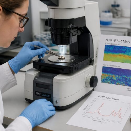
FTIR, on the other hand, identifies organic and inorganic compounds based on their molecular vibrations. It can identify binding media, varnishes, and organic pigments or dyes, which MSI cannot reliably do. This is a direct complement to MSI’s strength in pigment distribution mapping.
If MSI suggests an organic red dye, FTIR can help identify whether it is madder lake, cochineal, or another plant-based colorant. When combined, MSI maps the general location of pigment types, XRF identifies the metallic elements, and FTIR pinpoints the molecular structures of organic components and binders. This creates a multi-layered understanding.
This integrated approach allows conservators to build a robust material profile for an artwork, moving beyond mere surface appearances. It provides a comprehensive non-destructive art imaging strategy that covers a wide range of analytical questions. The synergy between these techniques offers unparalleled insights into artistic materials and techniques.
Case Examples Where Multispectral Imaging Changed Conservation Decisions
Multispectral imaging has profoundly impacted conservation decisions on numerous occasions, revealing hidden truths about famous artworks. One notable example involves Vincent van Gogh’s “Patch of Grass,” where MSI revealed an underlying portrait of a peasant woman beneath the landscape. This discovery changed how conservators approached cleaning and restoration, emphasizing the importance of preserving the hidden image.
Another compelling case involved a medieval illuminated manuscript, where multispectral pigment analysis helped differentiate between original pigments and later additions. Conservators had suspected extensive overpainting, but MSI precisely mapped the areas of original pigment versus later interventions. This information guided a more targeted and respectful conservation treatment, focusing on stabilizing the original layers.
In a Renaissance painting thought to be by a follower of Raphael, multispectral imaging pigment mapping non-invasive techniques revealed significant pentimenti and changes in composition. The sophisticated alterations suggested a master’s hand, prompting scholars to re-evaluate the attribution of the artwork. This led to a significant re-dating and re-contextualization of the piece within art history.
MSI was also instrumental in studying ancient Egyptian papyri, making faint and damaged hieroglyphs legible again. By capturing images in specific infrared bands, faded inks became visible, allowing Egyptologists to decipher previously unreadable texts. This non-destructive art imaging opened up new avenues for understanding ancient cultures.
A recent project on a pre-Columbian mural fragment used multispectral imaging to identify specific plant-based pigments used by indigenous artists. The pigment distribution mapping helped confirm trade routes and local resources, offering insights into the economic and cultural practices of the ancient civilization. These findings directly influenced how the mural was conserved and presented to the public.
These examples demonstrate that multispectral imaging is not just a diagnostic tool, but a catalyst for new discoveries and informed conservation. It allows us to see artworks through new eyes, respecting their complex histories and material realities. The insights gained from MSI prevent irreversible damage and ensure the longevity of cultural heritage.
The ability to visualize hidden layers and identify pigments non-invasively empowers conservators to make truly evidence-based decisions. It shifts conservation from a reactive process to a proactive, scientifically guided one. This method truly changes the game for preserving our shared artistic legacy.
From revealing an artist’s original intentions to uncovering the secrets of ancient texts, MSI offers a window into the past that was once impossible. It helps us understand the stories embedded within the very materials of an artwork. This technology continues to evolve, promising even more profound discoveries in the years to come.
Multispectral imaging pigment mapping non-invasive techniques are now an indispensable part of modern conservation science. They provide an incredible ability to study and understand historical pigments without ever touching them. This non-destructive art imaging approach is transforming the way we interact with and preserve our cultural treasures.
The detailed insights gained from multispectral pigment analysis allow conservators to develop highly specific and effective treatment plans. It ensures that interventions are minimal, reversible, and tailored to the unique material properties of each artwork. This precision helps maintain the authenticity and integrity of historical objects for future generations.
As technology advances, the capabilities of multispectral imaging will only grow, offering even finer spatial and spectral resolution. This means we can expect even more detailed pigment distribution mapping and deeper insights into complex artistic techniques. The future of art conservation looks promising with these powerful tools at our disposal.