Historical pigments hold stories within their chemical compositions and physical structures. Analyzing these pigments is essential for art conservation, authentication, and understanding the history of art itself. The advent of digital imaging microscopy pigments has transformed how we examine these tiny particles, offering unprecedented detail and accuracy.
This article explores the integration of digital imaging with microscopy techniques for the chemical analysis and conservation of historical pigments. We will discuss everything from image acquisition to image analysis, covering various microscopy techniques and their applications in pigment research and documentation.
Using digital imaging in microscopy is not merely about taking pretty pictures. It’s about extracting meaningful, quantifiable data that informs conservation strategies and helps us understand the materials and techniques used by artists of the past.
Integrating Digital Imaging with Microscopy
The integration of digital imaging with microscopy has revolutionized the field of pigment analysis. Traditional microscopy relied heavily on visual observation and manual recording, which were subjective and limited in scope.
Digital imaging enhances microscopy by allowing for the capture, storage, and manipulation of microscopic images. This offers several advantages, including improved image quality, the ability to perform quantitative image analysis, and the creation of detailed records for future reference.
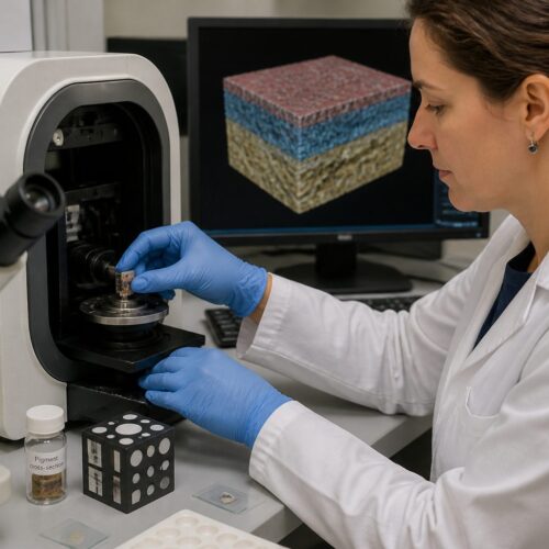
Different microscopy techniques can be combined with digital imaging to provide a comprehensive understanding of pigment characteristics. These techniques include optical microscopy, scanning electron microscopy (SEM), and transmission electron microscopy (TEM), each offering unique insights into pigment morphology and chemical composition.
Optical microscopy, for example, is useful for examining the color, shape, and size of pigment particles. SEM provides high-resolution images of the pigment surface, revealing details about texture and structure, while TEM allows for the examination of the internal structure of pigments at the nanometer scale.
The shift from analog to digital has also streamlined the process of sharing and collaborating on research findings. Digital images can be easily disseminated among researchers across the globe, fostering collaboration and accelerating the pace of discovery.
Furthermore, the ability to archive digital images ensures that valuable data is preserved for future generations of scientists and art historians. This long-term accessibility is crucial for longitudinal studies and for re-evaluating past findings in light of new knowledge and technologies.
The integration extends beyond image capture to include sophisticated software tools designed for image analysis and data processing. These tools enable researchers to quantify various aspects of pigment characteristics, such as particle size distribution, color intensity, and spatial arrangement.
By combining these quantitative data with other analytical techniques, such as spectroscopy and chromatography, a more complete picture of pigment composition and behavior can be obtained. This holistic approach is essential for addressing complex questions in art history and conservation science.
Camera Settings and Image Acquisition Techniques
Acquiring high-quality digital images in microscopy requires careful attention to camera settings and image acquisition techniques. The choice of camera is crucial, with options ranging from basic digital cameras to specialized scientific cameras designed for microscopy.
Key camera settings include resolution, exposure time, and gain, each of which affects the quality and information content of the image. High resolution is essential for capturing fine details, while appropriate exposure time and gain settings ensure that the image is neither under- nor overexposed.
Proper illumination is also critical for successful image acquisition. Different illumination techniques, such as brightfield, darkfield, and polarized light microscopy, can reveal different aspects of pigment structure and composition.
Brightfield illumination is the most common technique, providing a general view of the sample. Darkfield illumination enhances contrast by scattering light away from the objective lens, making it useful for visualizing small particles and surface features. Polarized light microscopy is particularly useful for identifying crystalline materials, which exhibit characteristic birefringence patterns.
Beyond the basic settings, understanding the nuances of color balance and white balance is paramount for accurate color representation. Incorrect color balance can lead to misinterpretations of pigment hues and shades, affecting the overall analysis.
Furthermore, the objective lens used in microscopy plays a critical role in image quality. Different objective lenses offer varying levels of magnification and numerical aperture, which affects the resolution and depth of field of the image.
Selecting the appropriate objective lens is essential for capturing the desired level of detail and for minimizing optical aberrations that can distort the image. Careful calibration of the microscope and camera system is also crucial for ensuring accurate measurements and reproducible results.
Advanced techniques like confocal microscopy can be employed to obtain optical sections of the sample, allowing for three-dimensional reconstruction of pigment structures. This is particularly useful for studying the layering and distribution of pigments within a complex matrix, such as a painting.
Image Processing and Enhancement Methods
Once digital images have been acquired, image processing and enhancement methods can be used to improve their quality and extract additional information. Image processing techniques include noise reduction, contrast enhancement, and sharpening, all of which can help to reveal subtle details in the image.
These techniques can be applied using a variety of software packages, such as ImageJ, Photoshop, and specialized microscopy software. Each software offers a range of tools for image manipulation and analysis, allowing researchers to optimize their images for specific purposes.
| Technique | Description | Application |
|---|---|---|
| Noise Reduction | Reduces random variations in pixel intensity | Improving image clarity |
| Contrast Enhancement | Adjusts the range of pixel intensities | Highlighting subtle features |
| Sharpening | Increases the apparent sharpness of edges | Enhancing fine details |
| Color Correction | Adjusts color balance | Ensuring accurate color representation |
It is important to note that image processing should be performed carefully to avoid introducing artifacts or misrepresenting the original data. The goal is to enhance the image without altering the underlying information, ensuring that any subsequent analysis is based on accurate data.
Furthermore, understanding the limitations of each image processing technique is crucial for interpreting the results accurately. Over-sharpening, for instance, can create artificial edges that are not actually present in the original sample.
Similarly, excessive noise reduction can blur fine details and obscure subtle features. A balanced approach is essential, where the goal is to improve image quality without compromising the integrity of the data.
Advanced image processing techniques, such as deconvolution, can be used to remove blurring caused by optical aberrations in the microscope. This can significantly improve the resolution and clarity of the image, allowing for the visualization of finer details.
Another important aspect of image processing is the use of appropriate color spaces and color management techniques. Different color spaces, such as RGB and CMYK, represent colors in different ways, and it is important to choose the appropriate color space for the specific application.
Analyzing Pigment Morphology and Distribution
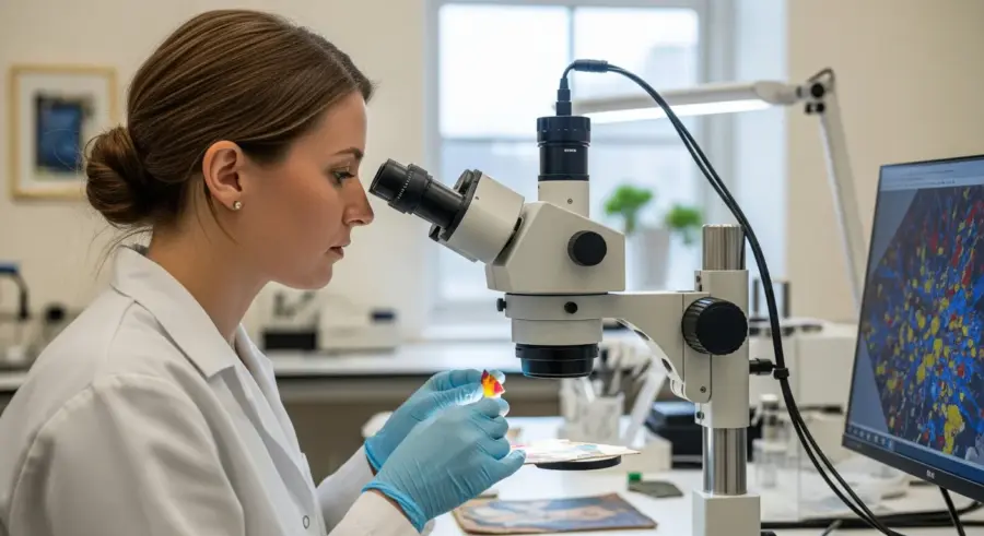
Digital imaging in microscopy enables detailed analysis of pigment morphology and distribution. Pigment morphology refers to the shape, size, and surface texture of individual pigment particles, while distribution refers to how these particles are arranged within a sample.
These characteristics can provide valuable information about the origin, processing, and application of the pigment. For example, the shape of a pigment particle can indicate whether it was produced naturally or synthetically, while the size distribution can reveal information about the grinding and mixing processes used to prepare the pigment.
Image analysis software can be used to quantify these morphological and distributional features. This involves segmenting the image to identify individual pigment particles, measuring their size and shape parameters, and analyzing their spatial arrangement.
By comparing these quantitative data with known standards, researchers can gain insights into the identity and history of the pigment. This information can be crucial for art historians and conservation scientists seeking to understand the materials and techniques used by artists of the past.
The analysis of pigment morphology can also reveal information about the degradation processes that have affected the pigment over time. For example, the presence of cracks or fissures on the pigment surface can indicate that the pigment has been exposed to environmental stress.
The distribution of pigments within a sample can also provide clues about the artist’s technique and the layering of different pigments. Analyzing the spatial relationships between different pigments can reveal information about the artist’s color mixing strategies and the order in which different layers of paint were applied.
Advanced image analysis techniques, such as fractal analysis, can be used to quantify the complexity of pigment morphology and distribution. Fractal analysis can provide insights into the surface roughness and irregularity of pigment particles, which can be related to their origin and processing.
Furthermore, the use of stereology techniques can allow for the three-dimensional reconstruction of pigment distributions within a sample. This provides a more complete understanding of the spatial arrangement of pigments and their relationships to other materials in the artwork.
Spectroscopic Techniques Enhanced by Digital Imaging
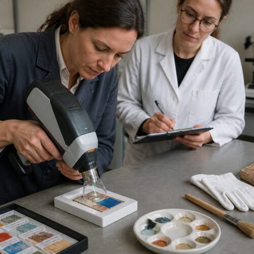
Spectroscopic techniques, such as Raman spectroscopy and X-ray fluorescence (XRF), can be enhanced by digital imaging to provide even more detailed information about pigment composition. Raman spectroscopy is a non-destructive technique that provides information about the molecular vibrations of a material, allowing for the identification of specific chemical compounds.
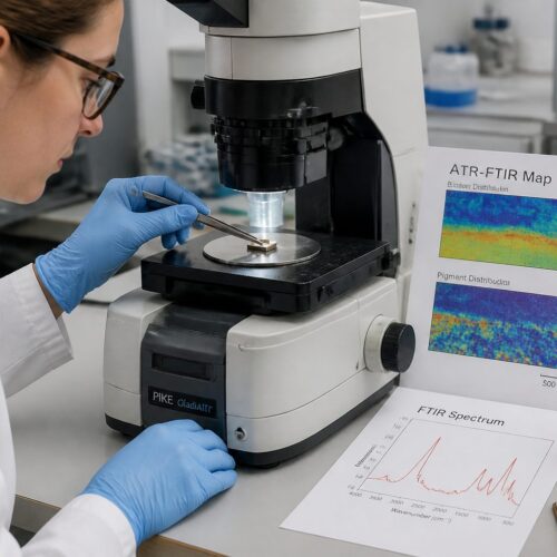
XRF is another non-destructive technique that provides information about the elemental composition of a material. By combining these techniques with digital imaging, researchers can create detailed maps of the chemical composition of a sample, revealing the distribution of different pigments and other materials within the artwork.
For example, Raman microscopy can be used to identify the specific crystalline forms of a pigment, such as anatase or rutile in the case of titanium dioxide. XRF mapping can reveal the presence of trace elements, which can provide clues about the origin of the pigment.
The combination of digital imaging and spectroscopic techniques offers a powerful approach to pigment analysis. It provides both visual and chemical information, allowing for a more complete understanding of the materials used in historical artworks.
The spatial resolution of spectroscopic techniques can be significantly enhanced by integrating them with digital imaging microscopy. This allows for the precise targeting of specific areas of interest within the sample, enabling the acquisition of detailed chemical information at the microscopic level.
For instance, Raman mapping can be used to identify the distribution of different pigments within a complex paint mixture, revealing the artist’s color mixing strategies. XRF mapping can be used to identify the presence of corrosion products or other degradation materials on the pigment surface.
The combination of digital imaging and spectroscopic techniques also allows for the creation of multi-modal datasets, which can be analyzed using advanced data fusion techniques. This involves combining information from different sources to create a more complete and integrated understanding of the sample.
For example, data from optical microscopy, Raman spectroscopy, and XRF can be combined to create a three-dimensional map of the pigment composition and distribution within a painting. This provides a powerful tool for understanding the materials and techniques used by artists and for assessing the condition of artworks.
Documenting Microscopic Findings with Digital Images
Documenting microscopic findings with digital images is essential for preserving and sharing research results. Digital images provide a permanent record of the microscopic appearance of pigments, allowing researchers to revisit their observations and share them with others.
Proper documentation includes not only the images themselves but also detailed metadata about the sample, the microscopy technique used, and the image acquisition parameters. This metadata ensures that the images can be interpreted correctly and that the research can be reproduced by others.
- Clearly label all images with sample information
- Include scale bars to indicate magnification
- Document the microscopy technique used
- Record camera settings and image acquisition parameters
- Store images in a standard, lossless format
In addition to still images, time-lapse microscopy can be used to document changes in pigment structure or composition over time. This can be particularly useful for studying the effects of conservation treatments or environmental factors on pigment stability.
Maintaining a detailed laboratory notebook is crucial for documenting all aspects of the research process. This notebook should include information about sample preparation, microscopy techniques, image processing methods, and data analysis procedures.
The use of standardized file naming conventions and data management systems is also essential for ensuring the long-term accessibility and usability of digital images. This includes using consistent naming schemes for image files and storing images in a secure and organized manner.
Furthermore, it is important to adhere to ethical guidelines for image manipulation and presentation. Images should be processed and enhanced in a manner that is consistent with scientific integrity and that does not misrepresent the original data.
The use of open-source software and data repositories can also promote transparency and reproducibility in pigment analysis research. This allows other researchers to access and analyze the data, verifying the results and building upon the findings.
Applications of Digital Imaging in Pigment Analysis
Digital imaging in microscopy has a wide range of applications in pigment analysis and conservation. One key application is the identification of pigments in historical artworks, which can provide valuable information about the artist’s materials and techniques.
By comparing the microscopic appearance and chemical composition of pigments in a painting with known standards, researchers can determine the identity of the pigments used. This information can be used to authenticate artworks, to understand the artist’s working methods, and to inform conservation strategies.
Another important application is the assessment of pigment degradation. Pigments can degrade over time due to exposure to light, moisture, and other environmental factors. Digital imaging can be used to monitor these changes, allowing conservation scientists to assess the condition of pigments and to develop appropriate conservation treatments.
For example, digital imaging can be used to track the fading of pigments, the formation of cracks or fissures, and the growth of microorganisms on the pigment surface. This information can be used to develop preventive conservation strategies, such as controlling the lighting and humidity in museum environments.
Digital imaging also plays a crucial role in the development of new conservation treatments. By visualizing the effects of different treatments on pigment structure and composition, conservation scientists can optimize their methods and minimize the risk of damage to the artwork.
Furthermore, digital imaging can be used to create virtual reconstructions of damaged or deteriorated artworks. This allows researchers to visualize the original appearance of the artwork and to understand how it has changed over time.
The application extends to the analysis of pigments in archaeological artifacts, providing insights into ancient technologies and trade routes. Identifying the pigments used in ceramics, textiles, and other artifacts can reveal information about the materials and techniques used by ancient cultures.
Moreover, digital imaging can be used to study the interaction between pigments and other materials in artworks, such as binders and varnishes. This can provide insights into the long-term stability of artworks and the factors that contribute to their degradation.
Case Studies: Digital Imaging in Action
Several case studies demonstrate the power of digital imaging in pigment analysis. One example involves the study of pigments in the paintings of Vincent van Gogh. Using digital imaging microscopy pigments, researchers have been able to identify the specific pigments used by van Gogh and to understand how these pigments have degraded over time.
This research has revealed that some of van Gogh’s favorite pigments, such as chrome yellow, are susceptible to light-induced degradation. As a result, museums have taken steps to protect van Gogh’s paintings from excessive light exposure.
Another case study involves the analysis of pigments in ancient Egyptian artifacts. Digital imaging has been used to identify the pigments used in wall paintings, sarcophagi, and other objects, providing insights into the materials and techniques used by ancient Egyptian artists.
This research has also revealed information about the trade routes and cultural exchanges that existed in ancient times, as some of the pigments used in Egyptian artifacts were imported from distant lands.
The study of the Dead Sea Scrolls has also benefited from digital imaging techniques. Researchers have used digital imaging to analyze the inks used to write the scrolls, providing insights into their composition and origin.
Digital imaging has also been used to analyze the pigments used in medieval illuminated manuscripts. This research has revealed information about the materials and techniques used by medieval artists and the sources of their pigments.
Another compelling case involves the investigation of Renaissance paintings, where digital imaging has helped uncover hidden layers and underdrawings beneath the visible surface. This provides valuable insights into the creative process of Renaissance masters.
These case studies highlight the diverse applications of digital imaging in pigment analysis and demonstrate its importance for understanding the history of art and culture.
Challenges and Future Directions
While digital imaging has greatly advanced the field of pigment analysis, several challenges remain. One challenge is the need for standardized protocols for image acquisition and analysis. Different researchers may use different techniques and software, making it difficult to compare results across studies.
Another challenge is the development of more sophisticated image analysis algorithms. Current algorithms are often limited in their ability to segment and quantify complex pigment mixtures, requiring manual intervention by the researcher.
Future directions in digital imaging for pigment analysis include the development of artificial intelligence (AI) and machine learning (ML) algorithms. These algorithms could automate the image analysis process, allowing for the rapid and accurate analysis of large datasets.
AI and ML could also be used to predict the degradation of pigments over time, based on their microscopic appearance and chemical composition. This would allow conservation scientists to develop more effective preventive conservation strategies.
The development of more user-friendly and accessible image analysis software is also a key priority. This would allow researchers with limited expertise in image processing to effectively analyze their data and contribute to the field.
Another important area of research is the development of new imaging techniques that can provide even more detailed information about pigment structure and composition. This includes techniques such as super-resolution microscopy and three-dimensional electron microscopy.
The integration of digital imaging with other analytical techniques, such as mass spectrometry and chromatography, is also a promising area of research. This would allow for the comprehensive characterization of pigments and other materials in artworks.
Addressing these challenges and pursuing these future directions will further enhance the power of digital imaging for pigment analysis and contribute to a deeper understanding of art history and conservation science.
Conclusion
Digital imaging in microscopy has transformed the field of pigment analysis, providing researchers with powerful tools for understanding the materials and techniques used by artists of the past. From image acquisition to image analysis, digital imaging offers unprecedented detail and accuracy, enabling new discoveries and informing conservation strategies.
As technology continues to advance, we can expect even more sophisticated imaging and analysis techniques to emerge, further enhancing our understanding of historical pigments. The future of pigment analysis is bright, with digital imaging playing a central role in unlocking the secrets of the past.
The integration of digital imaging not only enhances our ability to analyze pigments but also fosters greater collaboration and knowledge sharing within the art conservation and historical research communities. The accessibility of digital data promotes transparency and allows for the validation and refinement of research findings.
Ultimately, the insights gained through digital imaging techniques contribute to the preservation and appreciation of our cultural heritage. By understanding the materials and techniques used by artists of the past, we can develop more effective conservation strategies and ensure that these artworks are enjoyed by future generations.