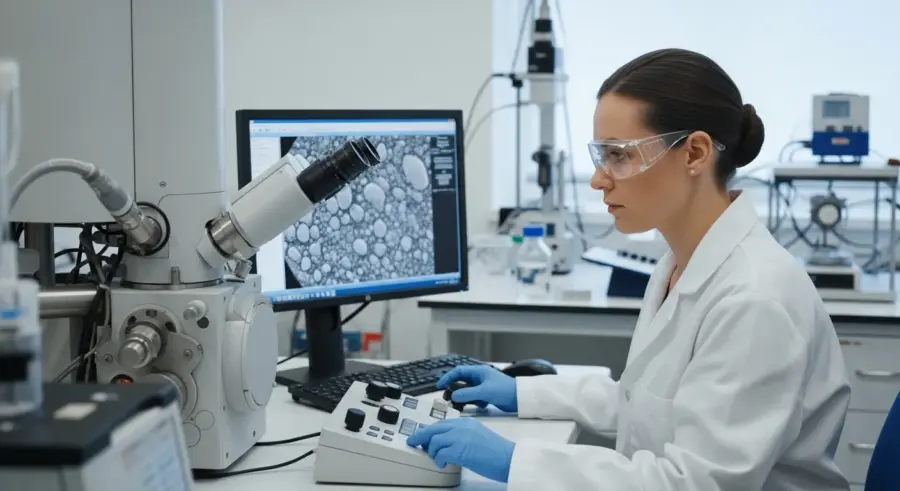
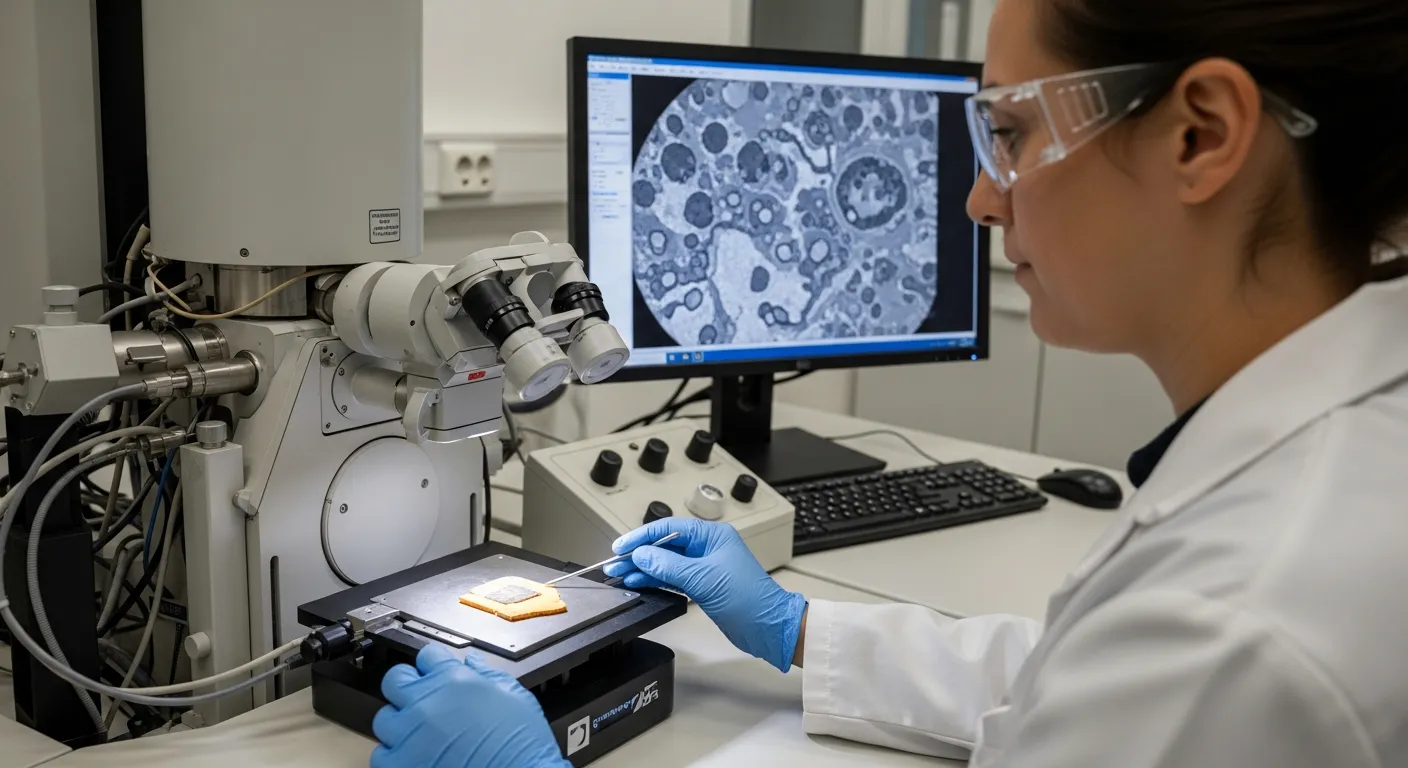
Scanning Electron Microscopy (SEM) has become an indispensable tool in the field of art conservation, especially when it comes to analyzing historical pigments. It offers a high-resolution imaging technique that allows us to examine the surface morphology and elemental composition of pigments with remarkable precision.
This detailed analysis is essential for understanding the materials used by artists, identifying degradation processes, and authenticating artworks. In this article, we will explore the principles behind Scanning Electron Microscopy, its application in pigment analysis, and its significance in the conservation of cultural heritage.
From sample preparation to data interpretation, we will cover the key aspects of using Scanning Electron Microscopy pigments to gain valuable insights into the world of historical art materials. We’ll also touch on how SEM imaging helps us understand the elemental composition and microstructure of these pigments, crucial for their preservation.
Principles of Scanning Electron Microscopy
Scanning Electron Microscopy utilizes a focused beam of electrons to scan the surface of a sample, providing detailed images at magnifications far beyond those achievable with optical microscopes. The interaction between the electron beam and the sample generates various signals, which are detected and used to create an image.
These signals include secondary electrons, backscattered electrons, and characteristic X-rays. Secondary electrons are low-energy electrons emitted from the sample’s surface and are used to create high-resolution images of the sample’s topography.
Backscattered electrons, on the other hand, are higher-energy electrons that are reflected or backscattered out of the sample. The intensity of backscattered electrons is strongly related to the atomic number of the elements in the sample, providing compositional information.
Characteristic X-rays are emitted when the electron beam displaces an inner-shell electron in an atom, and an outer-shell electron fills the vacancy. Each element emits X-rays with characteristic energies, allowing for elemental identification and quantification using Energy-Dispersive X-ray Spectroscopy (EDS).
The electron beam is generated by an electron gun, typically a tungsten filament or a field emission source. This beam is then focused by a series of electromagnetic lenses and apertures. These lenses precisely control the beam’s size and position as it scans across the sample.
The scanning pattern is usually a raster pattern, similar to that used in television screens. As the electron beam interacts with the sample, the emitted signals are detected by specialized detectors. These detectors convert the signals into electrical currents, which are then amplified and processed to create an image.
The magnification of the Scanning Electron Microscopy is determined by the ratio of the scanned area on the sample to the size of the display screen. By reducing the scanned area, the magnification can be increased. Modern Scanning Electron Microscopes can achieve magnifications of up to 1,000,000x.
Vacuum conditions are essential within the Scanning Electron Microscope to prevent scattering of the electron beam by air molecules. This ensures that the electron beam reaches the sample with minimal distortion. The vacuum also helps to prevent contamination of the sample surface.
Different detectors are used to capture the various signals produced by the electron beam interaction. Secondary electron detectors are usually of the Everhart-Thornley type, which uses a scintillator to convert electrons into photons. Backscattered electron detectors are typically solid-state detectors, which directly convert electrons into electrical signals.
The choice of detector depends on the type of information that is desired. For high-resolution imaging of surface topography, secondary electron detectors are preferred. For compositional imaging, backscattered electron detectors are more suitable.
The accelerating voltage of the electron beam is another important parameter that affects the image quality and the depth of penetration of the electron beam. Higher accelerating voltages result in greater penetration depth, which can be useful for analyzing subsurface features. However, higher voltages can also increase the risk of beam damage.
Sample Preparation for SEM Analysis of Pigments
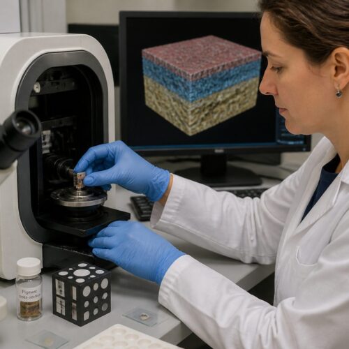
Proper sample preparation is crucial for obtaining accurate and reliable results from Scanning Electron Microscopy pigments analysis. Pigment samples are often in the form of powders, paint fragments, or cross-sections of painted layers.
For powder samples, a small amount of pigment is typically mounted on a conductive substrate, such as carbon tape or aluminum stubs. Paint fragments and cross-sections require more elaborate preparation to ensure a flat and stable surface for imaging.
These samples are often embedded in a resin, such as epoxy, and then polished to reveal the different layers of paint. Polishing is performed using progressively finer abrasives to achieve a smooth, scratch-free surface.
To enhance conductivity and prevent charge build-up during Scanning Electron Microscopy pigments analysis, samples are typically coated with a thin layer of conductive material, such as gold, platinum, or carbon. This coating is applied using a sputter coater, which deposits a uniform layer of the conductive material onto the sample’s surface.
When dealing with powder samples, it’s essential to ensure that the particles are well-dispersed on the conductive substrate. Agglomeration of particles can obscure their individual morphology and make it difficult to obtain accurate measurements. Sonication can be used to disperse the particles before mounting them on the substrate.
For paint fragments and cross-sections, the choice of embedding resin is critical. The resin should be chemically inert and should not react with the pigments or other components of the paint. It should also be hard enough to provide good support during polishing but not so hard that it damages the pigments.
The polishing process is a delicate balance between removing material to reveal the underlying layers and avoiding damage to the sample. Over-polishing can lead to the loss of fine details, while under-polishing can leave scratches and imperfections on the surface.
The thickness of the conductive coating is also an important consideration. A coating that is too thin may not provide adequate conductivity, leading to charge build-up and image distortion. A coating that is too thick can obscure the surface features of the sample.
Carbon coating is often preferred for EDS analysis because carbon is a light element and does not interfere with the detection of other elements. Gold and platinum coatings, on the other hand, can mask the signals from elements with similar X-ray energies.
In some cases, it may be necessary to use a focused ion beam (FIB) to prepare samples for Scanning Electron Microscopy. FIB milling can be used to create ultra-thin cross-sections or to remove specific areas of the sample to reveal underlying structures.
Analyzing Pigment Morphology with SEM
Scanning Electron Microscopy excels at revealing the morphology of pigment particles. The high magnification capabilities allow for detailed examination of particle size, shape, and surface texture.
This information can be used to differentiate between different pigment types and to assess the degree of pigment dispersion within a paint matrix. For example, natural pigments often exhibit irregular shapes and a wide range of particle sizes, while synthetic pigments tend to be more uniform.
| Pigment Type | Particle Shape | Particle Size | Surface Texture |
|---|---|---|---|
| Natural Ultramarine | Irregular | Variable (1-50 μm) | Rough, fractured |
| Synthetic Ultramarine | Rounded | Uniform (1-2 μm) | Smooth |
| Lead White | Acicular (needle-like) | Variable (1-10 μm) | Smooth to slightly rough |
| Vermilion | Rounded to angular | Variable (1-30 μm) | Smooth, sometimes faceted |
The table above illustrates typical differences in morphology between various pigments. Analyzing these characteristics helps in pigment identification and understanding the manufacturing processes used to create them.
The shape of pigment particles can provide clues about their origin and method of preparation. For instance, the acicular (needle-like) shape of lead white is a result of the chemical process used to produce it. Similarly, the rounded shape of synthetic ultramarine is due to the high-temperature calcination process.
Surface texture can also be a useful indicator of pigment type. Some pigments, such as natural ultramarine, have a rough, fractured surface due to the grinding process used to extract them from the mineral lapis lazuli. Other pigments, such as vermilion, may have a smooth, faceted surface due to the crystallization process.
The dispersion of pigments within a paint matrix can affect the optical properties of the paint. Poorly dispersed pigments can lead to uneven color and reduced gloss. Scanning Electron Microscopy can be used to assess the degree of pigment dispersion and to identify areas of agglomeration.
Quantitative analysis of particle size and shape can be performed using image analysis software. This allows for the creation of histograms and other statistical representations of the pigment morphology. These data can be used to compare different pigment samples and to track changes in pigment morphology over time.
It’s important to note that the morphology of pigments can be affected by degradation processes. For example, the surface of lead white can become corroded and pitted due to exposure to pollutants. Scanning Electron Microscopy can be used to identify these degradation features and to assess the extent of damage.
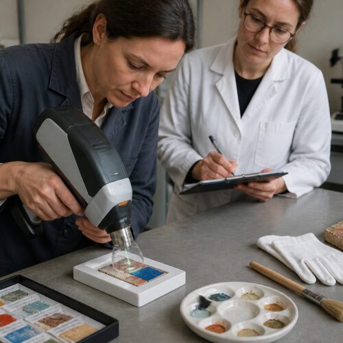
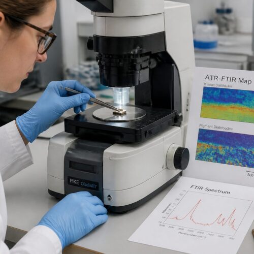
Using Energy-Dispersive X-ray Spectroscopy (EDS) for Elemental Analysis
Energy-Dispersive X-ray Spectroscopy (EDS) is often used in conjunction with Scanning Electron Microscopy to determine the elemental composition of pigments. When the electron beam interacts with the sample, it causes the emission of characteristic X-rays, which are then detected by the EDS detector.
By analyzing the energies and intensities of these X-rays, the types and amounts of elements present in the sample can be determined. This technique is particularly useful for identifying pigments based on their elemental fingerprints.
For example, the presence of mercury indicates the use of vermilion, while the presence of lead suggests lead white. EDS can also be used to identify impurities or additives in pigments, providing further information about their origin and manufacturing process.
Quantitative EDS analysis can provide the elemental ratios within a pigment, aiding in the identification of specific compounds. This is especially useful when dealing with complex pigments or mixtures of pigments.
The EDS detector measures the energy of the emitted X-rays and counts the number of X-rays at each energy level. The resulting spectrum shows peaks at specific energies that correspond to the elements present in the sample. The height of each peak is proportional to the concentration of the corresponding element.
Qualitative EDS analysis involves identifying the elements present in the sample based on the positions of the peaks in the spectrum. Quantitative EDS analysis involves determining the concentrations of the elements by comparing the peak heights to those of known standards. This requires careful calibration of the EDS system.
EDS analysis can be performed on individual pigment particles or on larger areas of the sample. Point analysis provides information about the elemental composition of a specific location. Mapping analysis creates a spatial distribution of the elements across the sample surface.
The accuracy of EDS analysis can be affected by several factors, including the sample preparation method, the accelerating voltage of the electron beam, and the presence of overlapping X-ray peaks. Careful attention to these factors is necessary to obtain reliable results.
EDS can be used to identify the presence of degradation products in pigments. For example, the formation of lead sulfates or lead chlorides on the surface of lead white can be detected by EDS. This information can be used to assess the condition of the artwork and to develop appropriate conservation treatments.
Identifying Pigment Degradation with SEM
Scanning Electron Microscopy is a valuable tool for studying pigment degradation in artworks. Changes in pigment morphology and elemental composition can indicate deterioration caused by environmental factors, such as light, humidity, and pollutants.
For instance, the degradation of lead white can result in the formation of lead carbonates or lead sulfides, which can be detected by EDS. SEM imaging can also reveal the formation of cracks, fissures, or other surface defects associated with pigment degradation.
- Chalking: Loss of binder and pigment from the surface
- Cracking: Formation of cracks due to stress or environmental changes
- Discoloration: Alteration of pigment color due to chemical reactions
- Efflorescence: Formation of salt deposits on the surface
- Loss: Complete disappearance of pigment layer
These degradation phenomena can significantly impact the appearance and structural integrity of artworks. Understanding these processes is crucial for developing effective conservation strategies.
The formation of lead carbonates on lead white is a common degradation process that occurs in the presence of carbon dioxide and moisture. The lead carbonate crystals can grow on the surface of the pigment, causing it to become dull and powdery.
Lead sulfides can form when lead white is exposed to sulfur-containing pollutants, such as hydrogen sulfide. Lead sulfide is black in color and can cause the pigment to darken. This darkening is often referred to as “lead darkening” or “sulfiding.”
Copper-containing pigments, such as azurite and malachite, can also undergo degradation in the presence of moisture and pollutants. These pigments can react with acids to form copper chlorides or copper sulfates, which are often green or blue in color. The formation of these degradation products can cause the pigment to become discolored and unstable.
The degradation of organic pigments is often more complex than that of inorganic pigments. Organic pigments can fade or change color due to exposure to light, heat, or pollutants. They can also be susceptible to microbial attack.
Scanning Electron Microscopy can be used to identify the presence of microorganisms on the surface of pigments. EDS can be used to identify the elemental composition of the microorganisms and their metabolic products. This information can be used to develop strategies to prevent or control microbial growth on artworks.
Case Studies: SEM in Pigment Identification
Several case studies demonstrate the power of Scanning Electron Microscopy in pigment identification and art conservation. In one study, SEM was used to analyze the pigments in a 17th-century Dutch painting.
The analysis revealed the presence of a rare copper-containing pigment, which had not been previously identified in Dutch paintings of that period. This discovery provided new insights into the artist’s palette and the trade of pigments in the 17th century.
Another study used Scanning Electron Microscopy to investigate the degradation of pigments in a Roman fresco. The analysis showed that the pigments had undergone significant chemical alteration due to exposure to moisture and pollutants.
The identification of these degradation products helped conservators develop a targeted conservation treatment to stabilize the fresco and prevent further deterioration. These examples show how Scanning Electron Microscopy pigments analysis can provide critical information for art historical research and conservation practice.
In the analysis of a medieval illuminated manuscript, Scanning Electron Microscopy with EDS was instrumental in identifying the presence of orpiment, a yellow arsenic sulfide pigment. Its use helped art historians understand the artistic techniques of the time and the origin of the materials.
The examination of a Van Gogh painting revealed the presence of zinc yellow, a pigment known to degrade over time and cause discoloration. The SEM images showed the altered morphology of the pigment particles, providing insight into the extent of the degradation.
Scanning Electron Microscopy has also been used to study the pigments in ancient Egyptian artifacts. Analysis of pigments from sarcophagi and tomb paintings has revealed valuable information about the materials used by the ancient Egyptians and their painting techniques.
A study of Renaissance paintings used Scanning Electron Microscopy to identify the layering techniques employed by the artists. The analysis of paint cross-sections revealed the sequence in which the different pigments were applied, providing insight into the artist’s working methods.
The use of Scanning Electron Microscopy in these case studies highlights its versatility and importance in art historical research and conservation. It allows researchers to gain a deeper understanding of the materials and techniques used by artists throughout history and to develop effective strategies for preserving our cultural heritage.
Applications in Forensic Analysis
Beyond art conservation, Scanning Electron Microscopy pigments analysis plays a role in forensic science. The ability to identify and compare trace amounts of pigments can be crucial in criminal investigations.
For example, pigment particles found on a suspect’s clothing can be compared to pigments from a crime scene, such as paint from a car or building. The unique morphology and elemental composition of pigments, as revealed by Scanning Electron Microscopy and EDS, can provide strong evidence linking a suspect to a crime.
Forensic analysis often involves analyzing small or degraded samples, making the high sensitivity and spatial resolution of Scanning Electron Microscopy particularly valuable. The technique can also be used to analyze pigments in counterfeit products, helping to identify the source and methods of production.
The application of Scanning Electron Microscopy in forensic science highlights its versatility and importance in various fields requiring detailed material analysis. It’s a powerful tool for identifying microscopic clues.
In cases involving hit-and-run accidents, Scanning Electron Microscopy can be used to compare paint fragments found on the victim’s clothing with the paint on the suspect’s vehicle. The identification of matching pigments can provide strong evidence linking the suspect to the crime.
Scanning Electron Microscopy can also be used to analyze pigments in counterfeit currency and documents. The identification of unusual or inconsistent pigments can help to detect forgeries and to trace the source of the counterfeit materials.
In cases involving arson, Scanning Electron Microscopy can be used to analyze the residues of accelerants and other materials used to start the fire. The identification of specific pigments can provide clues about the type of materials used and the origin of the fire.
The analysis of pigments in soil samples can also be useful in forensic investigations. Different types of soil contain different pigments, and the identification of these pigments can help to determine the origin of the soil and to link a suspect to a particular location.
The use of Scanning Electron Microscopy in forensic science is constantly evolving as new techniques and applications are developed. The ability to analyze microscopic particles with high precision and accuracy makes it an invaluable tool for law enforcement and criminal justice.
Challenges and Limitations of SEM Analysis
While Scanning Electron Microscopy is a powerful analytical technique, it is important to acknowledge its challenges and limitations. Sample preparation can be time-consuming and requires specialized equipment and expertise.
The need for a conductive coating can also alter the surface characteristics of the sample, potentially affecting the accuracy of morphological analysis. Furthermore, Scanning Electron Microscopy is a surface-sensitive technique, providing information only about the outermost layers of the sample.
This can be a limitation when analyzing layered structures, such as paint cross-sections, where information about the underlying layers is also needed. Beam damage is another concern, particularly for sensitive organic pigments.
High-energy electron beams can cause changes in the sample’s structure or composition, leading to inaccurate results. Careful optimization of imaging parameters is necessary to minimize beam damage and obtain reliable data.
Charge build-up on non-conductive samples can cause image distortion and artifacts. While conductive coatings can help to mitigate this problem, they can also obscure surface features and interfere with EDS analysis.
The vacuum environment required for Scanning Electron Microscopy can also be a limitation for some samples. Volatile samples can evaporate or decompose under vacuum, making it difficult to obtain accurate results. Environmental Scanning Electron Microscopy (ESEM) can be used to analyze some volatile samples, but it is not suitable for all materials.
The interpretation of Scanning Electron Microscopy images can be subjective and requires experience and expertise. It is important to carefully consider the imaging parameters and the sample preparation method when interpreting the results.
The cost of Scanning Electron Microscopy equipment and analysis can be a barrier for some researchers and institutions. The initial investment in equipment can be significant, and ongoing maintenance and operating costs can also be substantial.
Despite these challenges and limitations, Scanning Electron Microscopy remains a powerful and versatile analytical technique. Ongoing advancements in technology and techniques are continually expanding its capabilities and addressing some of its limitations.
Advancements in SEM Technology
Ongoing advancements in Scanning Electron Microscopy technology are continually expanding its capabilities and addressing some of its limitations. Environmental Scanning Electron Microscopy (ESEM) allows for the analysis of non-conductive samples without the need for a conductive coating.
This is achieved by maintaining a low-pressure gaseous environment within the sample chamber, which reduces charge build-up on the sample surface. Field Emission Scanning Electron Microscopy (FESEM) utilizes a field emission electron source, which provides a higher brightness and smaller probe size compared to conventional SEM.
This results in improved image resolution and signal-to-noise ratio, allowing for the examination of finer details. Cryo-Scanning Electron Microscopy (cryo-SEM) involves freezing the sample to cryogenic temperatures, which can help preserve its structure and prevent beam damage.
This technique is particularly useful for analyzing hydrated or volatile samples. These advancements are making Scanning Electron Microscopy an even more versatile and powerful tool for pigment analysis and art conservation.
Focused Ion Beam (FIB) Scanning Electron Microscopy is another advanced technique that combines the capabilities of Scanning Electron Microscopy with FIB milling. This allows for the precise removal of material from the sample, enabling the creation of cross-sections and the analysis of subsurface features.
Transmission Scanning Electron Microscopy (TSEM) combines the principles of Scanning Electron Microscopy and Transmission Electron Microscopy (TEM). This technique allows for the analysis of ultra-thin samples with high resolution and contrast.
The development of new detectors and imaging modes is also expanding the capabilities of Scanning Electron Microscopy. For example, electron backscatter diffraction (EBSD) can be used to determine the crystallographic orientation of materials.
Advancements in software and image processing techniques are also improving the accuracy and efficiency of Scanning Electron Microscopy analysis. Automated image analysis algorithms can be used to quantify particle size, shape, and distribution.
These advancements are making Scanning Electron Microscopy an increasingly powerful tool for a wide range of applications, including materials science, nanotechnology, and biology. The ongoing development of new technologies and techniques will continue to expand the capabilities of Scanning Electron Microscopy in the future.
Conclusion
Scanning Electron Microscopy is an invaluable technique for the chemical analysis and conservation of historical pigments. Its high-resolution imaging and elemental analysis capabilities provide detailed insights into pigment morphology, composition, and degradation.
By understanding these aspects, conservators and art historians can gain a deeper appreciation of the materials and techniques used by artists throughout history. Scanning Electron Microscopy pigments analysis also aids in the development of effective conservation strategies to preserve our cultural heritage for future generations.
As Scanning Electron Microscopy technology continues to advance, its role in pigment analysis and art conservation will only become more significant. The ability to visualize and analyze pigments at the micro and nano-scale is essential for understanding the complexities of art materials and their behavior over time.
From authenticating artworks to identifying degradation mechanisms, Scanning Electron Microscopy provides crucial information for preserving our artistic legacy. Its applications extend beyond art, proving its worth in forensic science too.
The combination of high-resolution imaging and elemental analysis makes Scanning Electron Microscopy a unique and powerful tool. It provides information that cannot be obtained by other analytical techniques. As our understanding of art materials and degradation processes continues to grow, Scanning Electron Microscopy will play an increasingly important role in preserving our cultural heritage.
The future of Scanning Electron Microscopy in pigment analysis and art conservation is bright. With ongoing advancements in technology and techniques, we can expect to see even more sophisticated and powerful applications of this technique in the years to come.
The development of new detectors, imaging modes, and software tools will enable us to gain even deeper insights into the complexities of art materials and their behavior over time. This will ultimately lead to more effective conservation strategies and a greater appreciation of our artistic legacy.
Scanning Electron Microscopy is not just a tool for scientists and conservators; it is also a tool for art historians and the general public. By providing detailed images and information about art materials, it can help us to better understand and appreciate the works of art that have shaped our culture.
The insights gained from Scanning Electron Microscopy analysis can be used to educate the public about art materials and conservation issues. This can help to raise awareness of the importance of preserving our cultural heritage for future generations.