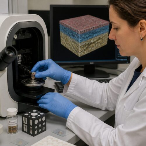
Art conservation relies heavily on understanding the materials that make up historical objects. Identifying the pigments used by artists is a critical aspect of this process. Polarized light microscopy pigments (PLM) offers a powerful and non-destructive method for analyzing these materials.
By examining the optical properties of pigment particles under polarized light, conservators can gain valuable insights into their composition and origin. This information is crucial for making informed decisions about the preservation and restoration of cultural heritage.
This article will explore the principles of polarized light microscopy pigments, its applications in art conservation, and the advantages and limitations of this technique. We will also discuss sample preparation methods and present case studies that demonstrate the effectiveness of PLM in pigment analysis.
Understanding Polarized Light
Polarized light is light that has been filtered so that its waves vibrate in a single plane. Ordinary light, such as that from the sun or a light bulb, vibrates in all directions perpendicular to its direction of travel.
When this light passes through a polarizing filter, only the light waves vibrating in a specific direction are allowed to pass through. This results in polarized light, which has unique properties that can be used to analyze anisotropic materials like many pigments.
The interaction of polarized light with anisotropic materials, those having optical properties that vary with direction, is the foundation of polarized light microscopy pigments. These materials can split a beam of polarized light into two rays that travel at different speeds and in different directions.
This phenomenon, known as birefringence, is a key characteristic used to identify different pigments. Birefringence is the numerical difference between the two refractive indexes.
Understanding the nature of light is paramount to grasping the principles behind PLM. Light, in its simplest form, is electromagnetic radiation that propagates as waves.
These waves oscillate in all directions perpendicular to their direction of travel. Polarization restricts these oscillations to a single plane, creating light with distinct properties.
This manipulation of light allows us to probe the internal structure and optical characteristics of materials at a microscopic level. The use of polarized light reveals details that would otherwise be invisible under normal illumination.
The polarizer, a key component in PLM, is responsible for creating this polarized light. It acts as a filter, selectively allowing light waves vibrating in a specific plane to pass through.
The resulting polarized light then interacts with the sample, providing valuable information about its composition and structure. The degree of polarization can be carefully controlled to optimize the analysis.
Birefringence, the splitting of light into two rays, is a direct consequence of the anisotropic nature of many pigments. This property is crucial for distinguishing between different pigment types.
The difference in refractive indices between the two rays provides a quantitative measure of birefringence. This value can be used to identify the pigment, often in conjunction with other optical properties.
The colors observed under polarized light are a result of the interference between these two rays. The thickness of the sample and the degree of birefringence influence the resulting color.
These interference colors are highly characteristic of specific pigments, making them a valuable tool for identification. Skilled conservators can use these colors to quickly narrow down the possibilities.
Setting Up a Polarized Light Microscope
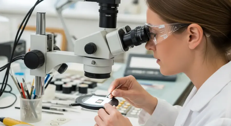
A polarized light microscope is a standard light microscope with additional components that allow for the observation of samples under polarized light. The main components include a polarizer, an analyzer, and a rotating stage.
The polarizer is placed below the sample and filters the light, producing polarized light that illuminates the specimen. The analyzer is placed above the sample, between the objective lens and the eyepiece, and it is also a polarizing filter.
The analyzer is typically oriented perpendicular to the polarizer, meaning that no light will pass through when there is no sample on the stage, resulting in a dark field of view. When an anisotropic sample is placed on the rotating stage, it interacts with the polarized light and alters its direction of vibration.
This change allows some of the light to pass through the analyzer, creating bright and colorful interference patterns that can be observed through the eyepiece. The rotating stage allows the sample to be rotated, changing the orientation of the pigment particles relative to the polarized light.
The proper alignment of the microscope components is critical for accurate results. The polarizer and analyzer must be precisely oriented perpendicular to each other, a condition known as crossed polars.
This ensures that only light that has been altered by the sample will pass through the analyzer. Any misalignment can lead to inaccurate observations and misidentification of pigments.
The objectives used in polarized light microscopy should be strain-free. Strain in the objective lens can introduce artificial birefringence, which can interfere with the analysis.
Special objectives designed for polarized light microscopy are available to minimize this effect. These objectives are carefully manufactured to ensure that they do not introduce any unwanted polarization effects.
The rotating stage is an essential feature of a polarized light microscope. It allows the sample to be rotated in the plane of the stage, changing the orientation of the pigment particles relative to the polarized light.
This rotation is crucial for determining the extinction position of the pigment particles. The extinction position is the orientation at which the particle appears darkest under crossed polars.
The rotating stage should be smooth and precise, allowing for accurate measurement of the extinction angle. A calibrated scale on the stage allows for precise determination of the angle.
The light source used in polarized light microscopy should be stable and provide even illumination. A halogen lamp is a common choice, but LED light sources are becoming increasingly popular due to their energy efficiency and long lifespan.
The intensity of the light source should be adjustable to optimize the contrast and visibility of the sample. Proper illumination is essential for obtaining clear and accurate images.
Identifying Pigments Based on Optical Properties
Polarized light microscopy pigments relies on the unique optical properties of pigments to identify them. These properties include birefringence, extinction position, and refractive index.
By carefully observing these characteristics under polarized light, conservators can differentiate between various pigments and gain insights into their composition. Birefringence, as mentioned earlier, is the splitting of a light beam into two rays with different refractive indices.
| Pigment | Birefringence | Extinction | Refractive Index |
|---|---|---|---|
| Ultramarine | Weak | Oblique | 1.50-1.52 |
| Malachite | Strong | Parallel | 1.65-1.91 |
| Vermilion | Very Strong | Parallel | 2.85-3.20 |
| Lead White | Weak | Parallel | 1.94-2.09 |
Extinction position refers to the orientation of a pigment particle when it appears darkest under crossed polars. Refractive index is a measure of how much light is bent when it passes from one medium to another.
The combination of these optical properties provides a unique “fingerprint” for each pigment. Skilled microscopists can use these fingerprints to identify pigments with a high degree of accuracy.
Birefringence is not just a simple on/off property; it varies in strength depending on the pigment. Some pigments exhibit strong birefringence, producing bright and vibrant interference colors.
Others exhibit weak birefringence, resulting in faint or subtle colors. The strength of birefringence is related to the degree of anisotropy in the pigment’s crystal structure.
The extinction position is another crucial characteristic for pigment identification. It is the angle at which the pigment particle appears completely dark under crossed polars.
The extinction position is related to the orientation of the pigment’s crystal axes relative to the polarized light. Pigments can exhibit parallel, oblique, or symmetrical extinction.
Refractive index is a fundamental property of a material that describes how much light bends when it passes through it. It is the ratio of the speed of light in a vacuum to the speed of light in the material.
Refractive index is usually determined by immersing the pigment particles in liquids of known refractive indices. When the refractive index of the liquid matches that of the pigment, the particle will disappear.
This technique, known as the Becke line method, allows for precise determination of the refractive index. The Becke line is a bright halo that appears around the particle when the refractive indices are mismatched.
By carefully observing the Becke line as the microscope is focused, the refractive index of the pigment can be determined. This is a critical step in identifying the pigment.
Preparing Samples for Polarized Light Microscopy
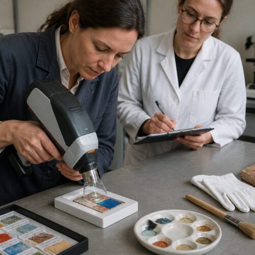
Proper sample preparation is essential for obtaining accurate and reliable results with polarized light microscopy pigments. The goal is to isolate individual pigment particles and mount them in a way that allows for optimal observation under the microscope.
One common method involves gently scraping a small amount of pigment from the artwork and dispersing it in a refractive index liquid on a glass slide. The refractive index liquid is chosen to have a refractive index close to that of the pigment being analyzed.
This helps to reduce the contrast between the pigment particles and the surrounding medium, making it easier to observe their optical properties. It is important to use a small amount of pigment to avoid overcrowding the slide, which can make it difficult to distinguish individual particles.
The sample should be evenly dispersed in the refractive index liquid and covered with a coverslip to protect the objective lens of the microscope. Another method involves embedding a small sample of the artwork in a resin, such as epoxy or acrylic.
When scraping pigment samples, it’s crucial to use a clean and sharp tool to avoid contamination. A micro-spatula or a fine needle can be used to carefully remove a small amount of pigment from the artwork.
The area from which the sample is taken should be carefully chosen to minimize damage to the artwork. Often, samples are taken from areas that are already damaged or have been previously restored.
The choice of refractive index liquid is critical for obtaining optimal results. A range of refractive index liquids are available, with refractive indices ranging from 1.40 to 1.70 or higher.
The refractive index of the liquid should be close to that of the pigment being analyzed to minimize contrast and enhance visibility. Databases and reference materials provide information on the refractive indices of various pigments.
When embedding samples in resin, it’s important to use a resin that is transparent and has a low refractive index. Epoxy and acrylic resins are commonly used for this purpose.
The sample should be carefully oriented in the resin to allow for optimal viewing under the microscope. The resin block is then polished to create a smooth surface for observation.
For some samples, it may be necessary to use a microtome to create thin sections for analysis. A microtome is a precision instrument that can cut extremely thin slices of material.
Thin sections are particularly useful for analyzing layered structures, such as paint cross-sections. The thickness of the section should be optimized to allow for clear observation of the pigment layers.
Advantages and Limitations of PLM in Pigment Analysis
Polarized light microscopy pigments offers several advantages for pigment analysis in art conservation. It is a relatively simple and inexpensive technique that can be performed with readily available equipment.
It is also a non-destructive or micro-destructive method, requiring only a very small sample of pigment, which is crucial when dealing with valuable historical objects. PLM can provide valuable information about the composition, origin, and alteration of pigments.
- Relatively inexpensive
- Requires minimal sample
- Can be performed on-site
- Provides data about pigment alteration
- Aids in understanding artist technique
However, PLM also has some limitations. It requires a skilled operator with experience in identifying pigments based on their optical properties.
One of the key advantages of PLM is its ability to differentiate between pigments that may appear similar under normal light. The unique optical properties revealed under polarized light provide a powerful means of distinguishing between them.
This is particularly useful for identifying pigments that have been altered by aging or environmental factors. PLM can also provide information about the particle size and shape of the pigments.
The minimal sample requirement of PLM is a significant advantage when analyzing valuable artwork. Only a tiny amount of pigment is needed to obtain reliable results.
This minimizes the risk of damage to the artwork and allows for the analysis of even the smallest fragments of pigment. The portability of PLM equipment allows for on-site analysis of artwork.
This can be particularly useful for museums and galleries that are unable to transport artwork to a laboratory. On-site analysis can provide valuable information quickly and efficiently.
Despite its advantages, PLM also has some limitations. The identification of pigments based on optical properties requires a skilled and experienced operator.
The interpretation of the interference patterns and extinction positions can be challenging, especially for complex mixtures of pigments. The accuracy of PLM depends on the quality of the sample preparation.
Poorly prepared samples can lead to inaccurate results and misidentification of pigments. PLM is not always able to identify all pigments definitively.
Some pigments have similar optical properties, making it difficult to distinguish between them. In these cases, other analytical techniques may be necessary to confirm the identification.
Case Studies: PLM in Art Conservation
Polarized light microscopy pigments has been used in numerous case studies to solve problems related to art conservation. One example is the analysis of pigments in a 15th-century Italian panel painting.
PLM was used to identify the pigments used by the artist, which included ultramarine, vermilion, and lead white. This information helped conservators understand the artist’s technique and the materials used in the painting.
It also provided insights into the painting’s condition and the potential for future deterioration. In another case study, PLM was used to analyze the pigments in a collection of ancient Egyptian artifacts.
The analysis revealed the presence of several pigments, including Egyptian blue, malachite, and ochre. By comparing the pigments found in different artifacts, researchers were able to gain insights into the trade routes and artistic practices of ancient Egypt.
In the 15th-century Italian panel painting case, PLM not only identified the pigments but also revealed information about their quality and origin. The ultramarine, for example, was found to be of high quality, indicating that the artist had access to expensive materials.
The presence of lead white suggested that the artist used a traditional painting technique. This information helped conservators to better understand the painting’s construction and to develop appropriate conservation strategies.
The analysis of the ancient Egyptian artifacts provided insights into the pigments used in different periods. The presence of Egyptian blue, a synthetic pigment, indicated that the artifacts were likely from the Roman period.
The identification of malachite and ochre, natural pigments, suggested that these pigments were used throughout Egyptian history. This information helped researchers to understand the evolution of artistic practices in ancient Egypt.
Another interesting case study involved the analysis of pigments in a collection of Chinese paintings. PLM was used to identify the pigments used in the paintings, which included azurite, cinnabar, and orpiment.
The analysis also revealed information about the layering of the pigments, which provided insights into the artist’s technique. The identification of specific pigments helped to determine the age and origin of the paintings.
In a case involving a suspected forgery, PLM was used to analyze the pigments in a painting attributed to a famous artist. The analysis revealed the presence of pigments that were not available during the artist’s lifetime.
This evidence, combined with other analytical data, confirmed that the painting was a forgery. PLM can be a powerful tool for detecting forgeries and authenticating artwork.
Conclusion
Polarized light microscopy pigments is a valuable tool for pigment analysis in art conservation. Its ability to provide information about the composition, origin, and alteration of pigments makes it an essential technique for conservators and art historians.
While PLM has some limitations, its advantages, such as its simplicity, low cost, and non-destructive nature, make it a powerful method for studying historical pigments. As technology advances, PLM continues to evolve, with new techniques and applications being developed to further enhance its capabilities.
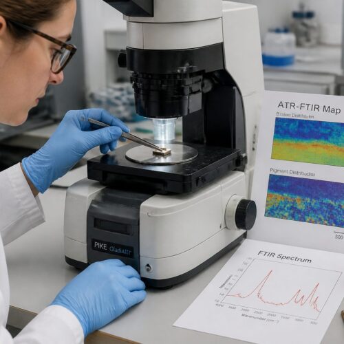
By combining PLM with other analytical techniques, such as X-ray diffraction and Raman spectroscopy, conservators can gain a more complete understanding of the materials used in historical objects. This knowledge is crucial for making informed decisions about their preservation and restoration, ensuring that these treasures are protected for future generations.
The ongoing research and development in the field of polarized light microscopy pigments promise to further refine our understanding of historical pigments and their role in art history. PLM’s contribution to conservation efforts will undoubtedly continue to grow.
The future of PLM in art conservation looks promising. New advancements in microscope technology and image analysis software are making it easier to acquire and interpret PLM data.
The development of new refractive index liquids and mounting media is also improving the quality of sample preparation. These advancements will further enhance the capabilities of PLM and make it an even more valuable tool for art conservators.
The integration of PLM with other analytical techniques is also becoming increasingly common. Combining PLM with techniques such as Raman spectroscopy and X-ray diffraction provides a more comprehensive understanding of the composition and structure of pigments.
This multi-analytical approach allows conservators to obtain a more complete picture of the materials used in historical objects. The training of skilled microscopists is essential for the continued success of PLM in art conservation.
Workshops and training programs are needed to ensure that conservators have the knowledge and skills necessary to perform accurate PLM analysis. The sharing of knowledge and expertise among conservators is also crucial.
Collaboration and communication can help to solve complex problems and advance the field of PLM in art conservation. The preservation of our cultural heritage is a shared responsibility.
By using techniques like polarized light microscopy pigments, we can gain a better understanding of the materials used in historical objects and make informed decisions about their preservation and restoration. This ensures that these treasures are protected for future generations to enjoy.