
The field of conservation has made significant strides with the advent of micro-CT scanning technology, particularly in the analysis of historical pigments, which are crucial in understanding the artistry of past generations. This innovative approach allows conservators to delve deep into the microstructure of paint layers without causing any damage to the artwork, thereby preserving the integrity of these invaluable pieces of cultural heritage.
The principles of micro-CT involve using X-rays to create high-resolution, three-dimensional images of an object’s internal structure, which is vital for thorough analysis. By capturing detailed images of the pigment layer microstructure, conservators can gain insights into the artwork’s history and condition, enabling them to make informed decisions about preservation and restoration techniques.
Micro-CT’s resolution capabilities are particularly impressive, often reaching sub-micron levels, which is crucial when it comes to analyzing fine details in paint stratigraphy. This high level of detail enables conservators to identify individual layers of paint, including underdrawings and ground layers, with unparalleled accuracy, allowing for a thorough understanding of the artist’s techniques and materials.
Understanding the principles of micro-CT is essential for effective application in conservation, as it lays the groundwork for its use in various analyses. This technology uses a rotating X-ray source and detector to capture multiple images, which are then reconstructed into a 3D model that reveals complex internal structures otherwise invisible to the naked eye.
The resolution of micro-CT scans can vary based on the equipment used, but many systems offer resolutions down to 1 micron, allowing for an incredibly detailed examination of artworks. Such precision makes it possible to visualize the complex layering of historical paints and their respective microstructures, which is vital for accurate conservation assessments.
Furthermore, the ability to manipulate the reconstructed images allows researchers to focus on specific areas of interest within the sample, enhancing the granularity of their analysis. This targeted approach not only enhances the overall understanding of the artwork’s composition and condition but also aids in developing tailored conservation strategies.
By using advanced algorithms, micro-CT can distinguish between different materials based on their density, an essential feature for conservation work. This capability is particularly useful in identifying various pigment types and binding media within the paint layers, thus informing conservators about the historical context and the techniques employed by the artist.
In summary, the principles of micro-CT provide a solid foundation for its application in the conservation of historical pigments, revolutionizing the field. The high-resolution capabilities of this technology enable a detailed analysis that was previously unattainable, allowing for a more profound understanding of the artworks’ intricacies.
The advent of three-dimensional layer data significantly alters the interpretation of paint stratigraphy, opening new avenues for art historical research and conservation practice. Traditionally, stratigraphy was examined in two dimensions, often leading to misinterpretations of layer order and composition that could misguide conservation efforts.
With 3D paint layer analysis, conservators can visualize the full extent of each layer, revealing their relationships in a more comprehensive manner that enhances historical understanding. This holistic view allows for greater accuracy in determining the techniques and materials used by the artist, fostering a deeper appreciation for their methodologies.
Moreover, 3D data facilitates the identification of subtle features, such as variations in layer thickness and the presence of intermediate layers, which are often critical to understanding an artwork’s creation. These details can provide vital clues about the artist’s methods and the artwork’s provenance, enriching the narrative surrounding each piece.
As a result, the analysis of paint layers can inform decisions related to conservation strategies, ensuring that interventions are both appropriate and effective. Understanding the original application methods enables conservators to preserve the integrity of the artwork while preventing further deterioration, thereby extending its lifespan for future generations.
In conclusion, three-dimensional layer data revolutionizes the way we interpret paint stratigraphy, offering a more nuanced understanding of historical artworks. This advancement enhances the overall conservation process and fosters a deeper appreciation for the complexities of artists’ techniques, ultimately enriching the cultural heritage we strive to protect.
When preparing samples for micro-CT conservation analysis, understanding the sample size requirements is crucial for achieving reliable results. The ideal sample size can vary depending on the specific objectives of the study, but generally, a minimum size of a few millimeters is recommended to ensure that the analysis is both effective and informative.
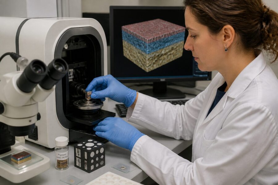
Additionally, proper sample preparation is vital to ensure high-quality imaging, as any imperfections in the sample can lead to artifacts in the scans. This includes careful cutting and mounting of the paint cross-section to maintain structural integrity and avoid compromising the layers that are to be analyzed.
| Sample Size (mm) | Preparation Method | Imaging Resolution (µm) |
|---|---|---|
| 1-2 | Polishing | 5 |
| 2-5 | Embedding in resin | 1 |
| 5-10 | Staining | 0.5 |
| 10+ | Multi-layer analysis | 0.1 |
In addition to size, the preservation of the paint layers during sample preparation is critical, as any mishandling can lead to irreversible damage that may compromise the analysis results. Ensuring that the layers remain intact and undisturbed is paramount for obtaining accurate data, making careful handling essential throughout the process.
Ultimately, careful consideration of sample size and preparation is essential for successful paint cross-section CT analysis, as these elements significantly influence the quality and accuracy of the resulting data. A well-prepared sample can provide invaluable insights into the artwork’s composition and condition, guiding conservators in their preservation efforts.
Micro-CT scanning provides an unparalleled opportunity to visualize crack networks, voids, and delamination within paint layers in three dimensions, offering vital information for conservation specialists. This insight is crucial for understanding the deterioration processes affecting historical artworks, enabling conservators to take proactive measures to prevent further damage.
Unlike traditional methods that may only offer a surface-level view, 3D imaging reveals complex internal structures that are often hidden from plain sight. This capability allows conservators to assess the extent of damage and plan appropriate conservation strategies effectively, ensuring that interventions are based on thorough analysis.
- Identification of crack networks
- Analysis of voids within layers
- Assessment of delamination
- Tracking changes over time
- Informing conservation strategies
Furthermore, visualizing these features in 3D can enhance the understanding of how environmental factors contribute to deterioration, providing a clearer picture of the conditions that affect artworks. By identifying specific problem areas within the paint structure, conservators can prioritize their intervention efforts to address the most critical concerns first.
Ultimately, the ability to visualize crack networks, voids, and delamination in 3D enriches the conservation process, making it more data-driven and effective. It provides essential data to formulate targeted restoration plans that preserve the integrity of the artwork while respecting its historical significance.
One of the most significant advantages of micro-CT scanning is the ability to correlate density maps with elemental composition, thereby enhancing the understanding of the materials used in historical pigments. This correlation allows researchers to infer the presence of different elements within the paint layers, which can shed light on both the materials employed and the techniques utilized by artists of the past.
By analyzing density variations, researchers can infer the presence of different elements within the paint layers, which is vital for identifying specific pigment types and understanding their historical context. This information not only assists in the identification of materials but also plays a critical role in appreciating the technological advancements and limitations of the time when the artwork was created.
Additionally, the correlation between density maps and elemental composition can provide insights into the degradation processes of the materials, which is essential for developing effective conservation strategies. Understanding how these materials react to environmental conditions and aging can guide conservators in making informed decisions regarding care and preservation.
This knowledge not only aids in the preservation of the artwork but also informs future restoration efforts, ensuring that interventions are appropriate and respectful of the original materials. As conservators work to maintain the integrity of historical pieces, this data proves invaluable in navigating the complexities of restoration.
In conclusion, correlating micro-CT density maps with elemental composition enhances the conservation of historical pigments significantly. This approach offers a deeper understanding of the materials and techniques employed by artists throughout history, enriching our appreciation of their contributions to the art world.
Micro-CT scanning has revolutionized the way conservators reveal underdrawing and ground texture non-invasively, offering a new tool for understanding the creative processes of artists. This capability allows for the examination of hidden layers without any physical intervention, preserving the artwork’s integrity while unveiling critical insights into its creation.
The non-invasive nature of this technology means that conservators can gain insights into the artist’s original intentions without risking damage to the artwork itself. This approach is particularly valuable for historical works where every detail counts, as it provides a glimpse into the artist’s thought process and techniques.
- Identification of underdrawings
- Mapping ground texture
- Analyzing compositional changes
- Assessing brushwork techniques
- Understanding artistic evolution
Moreover, the ability to visualize underdrawings and ground textures opens new avenues for understanding artistic practices and the evolution of style over time. This knowledge enriches our comprehension of historical artworks and provides context for their creation, enabling a more nuanced interpretation of the artist’s work.
In summary, revealing underdrawing and ground texture non-invasively through micro-CT scanning is a significant advancement in conservation technology. This technique enhances the understanding of artists’ methodologies while preserving the artwork’s historical integrity, ultimately contributing to the broader field of art conservation.
Effective analysis of micro-CT data relies heavily on powerful software tools for volume rendering and layer segmentation, which are essential for interpreting complex datasets. These tools enable researchers to manipulate and visualize intricate data effectively, facilitating a deeper understanding of the layers within artworks.
Advanced software allows for the segmentation of individual paint layers based on density differences, which is vital for conducting thorough analyses of the paint layer microstructure. This capability is essential for conservators seeking to unravel the complexities of historical pigments and their applications.
Furthermore, many software options offer user-friendly interfaces, making them accessible to conservators without extensive technical backgrounds or training. This democratization of technology enhances collaborative efforts in the field of conservation, allowing more professionals to engage with micro-CT data.
Additionally, the ability to share visualizations and findings through these platforms facilitates communication among researchers and conservators, promoting a collaborative approach to conservation efforts. This collaborative spirit fosters a broader understanding of historical pigments and their conservation, ultimately benefiting the entire field.
In conclusion, software tools for volume rendering and layer segmentation play a crucial role in micro-CT conservation analysis, enabling a more detailed interpretation of the data gathered. These tools empower conservators to extract valuable insights from complex datasets, ultimately enhancing their conservation efforts and the preservation of cultural heritage.
The application of micro-CT scanning in case studies has demonstrated its effectiveness in mapping panel painting structures and deterioration, providing a new lens through which to assess and preserve artworks. Through detailed imaging, conservators can assess the overall condition of the artwork and identify areas requiring intervention, ensuring that conservation efforts are both informed and targeted.
In one notable application, micro-CT revealed layering issues that traditional methods had overlooked, thereby highlighting the importance of this technology in modern conservation practice. This insight not only allowed for informed decisions on conservation treatments tailored to the specific needs of the artwork but also underscored the limitations of conventional techniques.
Moreover, the ability to visualize deterioration patterns in 3D provides a clearer understanding of how environmental factors have impacted the artwork over time, which is crucial for developing strategies to mitigate future damage. By linking deterioration patterns with environmental conditions, conservators can create more effective preservation plans that extend the life of the artwork.
Additionally, case studies often involve collaboration with researchers from various disciplines, fostering a multidisciplinary approach to conservation that enriches the overall effectiveness of conservation strategies. This collaborative effort enhances the depth of analysis and the range of solutions that can be applied to complex conservation challenges.
In summary, micro-CT scanning has proven invaluable in case applications related to panel painting structure and deterioration mapping, showcasing its vital role in modern art conservation. Its ability to provide detailed insights greatly enhances the conservation efforts undertaken by historians and conservators alike, ensuring that cultural heritage is preserved for future generations.
When comparing micro-CT with traditional SEM cross-section analysis, distinct advantages and limitations emerge that impact the choice of method for conservation studies. Micro-CT offers a non-destructive method that preserves the integrity of the artwork, whereas SEM typically requires physical sampling, which can lead to irreversible changes in the material being analyzed.
Additionally, micro-CT provides a more comprehensive view of the entire paint layer microstructure, allowing for a holistic understanding of the artwork’s composition. In contrast, SEM often focuses on surface analysis, potentially missing critical information about underlying layers that could be essential for a complete assessment.
Moreover, the three-dimensional data generated by micro-CT enables a better understanding of spatial relationships within the paint layers, which is particularly important for assessing complex deterioration processes. By visualizing the layers in three dimensions, conservators can identify patterns and issues that may not be evident in two-dimensional analyses.
On the other hand, SEM can provide elemental analysis with high spatial resolution, which is beneficial for identifying specific materials and compounds within the paint. However, the destructive nature of SEM sampling limits its applicability for valuable artworks that cannot afford to lose any material during the analysis process.
Ultimately, both methods have their place in the conservation of historical pigments, each offering unique advantages that can complement the other. Choosing the appropriate technique depends on the specific needs of the analysis and the condition of the artwork, ensuring that conservation efforts are both effective and respectful of the original materials.
As micro-CT technology continues to evolve, emerging miniaturized systems are becoming more accessible for smaller laboratories, which is a significant development in the field of conservation. These advancements are democratizing access to this powerful conservation tool, enabling more institutions to engage in detailed analyses of artworks.
Miniaturized CT systems offer comparable resolution capabilities to larger systems while requiring less space and lower operational costs, making them suitable for smaller labs that may not have the resources for larger, more expensive equipment. This accessibility encourages broader adoption of micro-CT scanning in the conservation community, leading to a more widespread understanding of historical materials.
Furthermore, these smaller systems are often designed with user-friendly interfaces, facilitating their integration into existing workflows and making them more approachable for conservators. This ease of use promotes collaboration among conservators, scientists, and researchers, fostering a community dedicated to the preservation of cultural heritage.
In addition to improving access, the development of these systems fosters innovation and research within the field, allowing smaller labs to participate in significant conservation projects. This participation contributes to the overall advancement of the discipline, enriching our understanding of historical artworks and their preservation needs.
In conclusion, the emergence of miniaturized CT systems presents exciting opportunities for smaller labs that seek to engage with cutting-edge conservation technology. This advancement enhances the accessibility of micro-CT conservation technology, ultimately benefiting the preservation of historical artworks and ensuring that vital cultural narratives are maintained.