X-ray fluorescence is a powerful analytical technique. It offers a unique way to examine the elemental composition of materials without causing damage.
This method is especially valuable in the field of art conservation. It allows us to study historical pigments and artworks in a non-destructive manner.
Using XRF spectroscopy, conservators and art historians can gain insights into the materials used by artists. This knowledge is crucial for authentication, preservation, and understanding artistic techniques.
Understanding the Principles of X-ray Fluorescence
X-ray Fluorescence (XRF) spectroscopy is based on the principle that individual elements emit characteristic X-rays when excited. This excitation is usually achieved by bombarding the sample with high-energy X-rays from an X-ray tube.
When an inner-shell electron is ejected, an electron from an outer shell fills the vacancy. This transition releases energy in the form of a fluorescent X-ray.
The energy of the emitted X-ray is specific to the element and the electron transition involved. By measuring the energies and intensities of these X-rays, we can determine the elemental composition of the sample.
The intensity of each X-ray line is proportional to the concentration of that element in the sample. This allows for both qualitative and quantitative elemental analysis.
The process begins with the emission of primary X-rays from the X-ray tube. These X-rays interact with the atoms in the sample being analyzed.
When a primary X-ray has sufficient energy, it can eject an electron from one of the atom’s inner electron shells, such as the K or L shell. This creates a vacancy in that electron shell.
The atom is now in an unstable state. An electron from a higher energy level (an outer shell) will drop down to fill the vacancy in the inner shell.
As the outer-shell electron moves to the inner shell, it releases energy equal to the difference in energy between the two electron shells. This energy is emitted as a fluorescent X-ray.
The energy of the fluorescent X-ray is characteristic of the element. Each element has a unique set of electron energy levels, and thus emits X-rays with specific energies.
A detector measures the energy and intensity of the emitted X-rays. The detector then generates a spectrum showing the intensity of X-rays at different energies.
The positions of the peaks in the spectrum correspond to the elements present in the sample. The intensities of the peaks are related to the concentrations of those elements.
By analyzing the XRF spectrum, we can identify the elements present in the sample and determine their concentrations. This provides valuable information about the composition of the material.
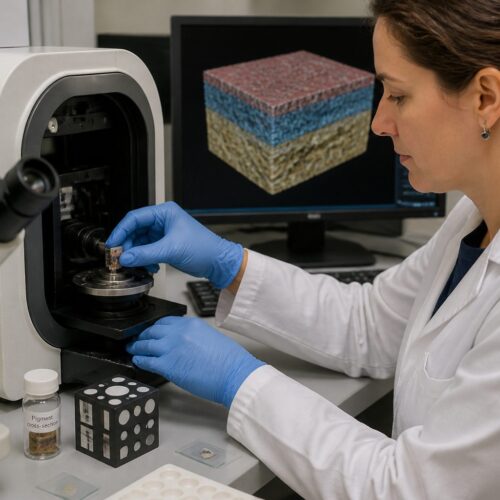
Preparing Samples for XRF Analysis
One of the major advantages of XRF is that minimal sample preparation is typically required. For many applications, the sample can be analyzed directly without any alteration.
However, some sample preparation steps may be necessary to ensure accurate and reliable results. These steps often depend on the type of sample and the specific analytical requirements.
For heterogeneous materials, grinding and homogenization can improve the accuracy of the analysis. This ensures that the X-ray beam interacts with a representative portion of the sample.
In some cases, samples may be pressed into pellets or fused with a flux to create a homogeneous glass disk. This is particularly useful for quantitative analysis of geological samples or materials with complex matrices.
The ideal sample for XRF analysis is flat, homogeneous, and of a suitable size to cover the area exposed to the X-ray beam. The sample should also be representative of the material being analyzed.
For powdered samples, grinding is often necessary to reduce particle size and improve homogeneity. A mortar and pestle or a ball mill can be used for this purpose.
After grinding, the powder can be pressed into a pellet using a hydraulic press. A binder, such as wax or cellulose, may be added to improve the mechanical strength of the pellet.
Alternatively, the powder can be fused with a flux, such as lithium borate, at high temperature. This creates a homogeneous glass disk that is ideal for quantitative analysis.
Liquid samples can also be analyzed by XRF. The liquid is typically placed in a special sample cup with a thin film window.
Care must be taken to ensure that the liquid is homogeneous and that there are no air bubbles in the sample cup. The thickness of the liquid layer must also be controlled to ensure accurate results.
For solid samples with irregular shapes, it may be necessary to use a sample holder to ensure that the sample is positioned correctly in the X-ray beam. The sample holder should be made of a material that does not interfere with the XRF analysis.
In some cases, it may be necessary to apply a thin film coating to the sample to improve its conductivity or to protect it from oxidation. The coating should be thin and uniform, and its composition should be known.
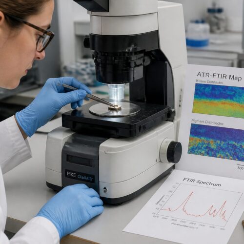
Interpreting XRF Spectra for Pigment Identification
Interpreting XRF spectra involves identifying the characteristic X-ray peaks associated with each element present in the sample. Each element emits a unique set of X-ray lines, allowing for unambiguous identification.
The XRF spectrum displays the intensity of X-rays as a function of their energy. By comparing the peak positions with known reference data, the elemental composition can be determined.
| Pigment Name | Primary Elements Detected | Typical X-ray Energies (keV) |
|---|---|---|
| Ultramarine | Na, Al, Si, S | 1.04 (Na), 1.49 (Al), 1.74 (Si), 2.31 (S) |
| Vermilion | Hg, S | 9.99 (S), 2.31 (Hg) |
| Lead White | Pb | 2.34 (Pb) |
| Egyptian Blue | Ca, Cu, Si | 3.69 (Ca), 8.04 (Cu), 1.74 (Si) |
Identifying the elements present allows for the determination of the pigments used in a painting or artifact. Knowing which elements correspond to which pigments is essential for accurate identification.
For example, the presence of mercury (Hg) and sulfur (S) strongly suggests the use of vermilion. Likewise, lead (Pb) indicates the presence of lead white.
The interpretation of XRF spectra requires a good understanding of X-ray physics and the characteristic X-ray energies of different elements. Reference databases and spectral analysis software are often used to aid in the identification of elements.
The XRF spectrum typically shows a series of peaks at specific energies. Each peak corresponds to a particular X-ray transition in a specific element.
The energy of the peak is determined by the difference in energy between the electron shells involved in the transition. The intensity of the peak is proportional to the concentration of the element in the sample.
In addition to the characteristic X-ray peaks, the spectrum may also contain background radiation. This background radiation can be caused by scattering of the primary X-rays or by other processes.
The background radiation can interfere with the identification of weak peaks. It is important to subtract the background from the spectrum before analyzing the peaks.
The presence of overlapping peaks can also complicate the interpretation of XRF spectra. Overlapping peaks occur when the X-ray energies of two or more elements are very close together.
In these cases, it may be necessary to use spectral deconvolution techniques to separate the overlapping peaks. Spectral deconvolution involves fitting mathematical functions to the peaks in the spectrum to determine their individual intensities.
The identification of pigments in artworks can be challenging due to the complex mixtures of pigments that are often used. Artists often mix different pigments to achieve the desired colors and effects.
In these cases, it is important to consider the possible combinations of pigments that could produce the observed XRF spectrum. Historical recipes and artistic techniques can provide valuable clues.
Advantages and Limitations of XRF in Art Conservation
XRF offers several advantages for the analysis of historical pigments. Its non-destructive nature is particularly valuable when dealing with rare or fragile artworks.
The technique is relatively fast and requires minimal sample preparation. It can be used to analyze a wide range of materials, including pigments, metals, and ceramics.
However, XRF also has some limitations. It is primarily a surface technique, with limited penetration depth.
Light elements, such as hydrogen, carbon, and oxygen, are difficult to detect with XRF. The spatial resolution of XRF is also limited compared to other microanalytical techniques.
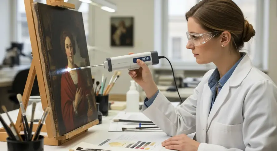
One of the main advantages of XRF is its ability to analyze materials in situ. This means that the analysis can be performed directly on the artwork without the need to remove a sample.
This is particularly important for fragile or valuable artworks that cannot be easily moved or sampled. Portable XRF instruments are available that can be used in museums or archaeological sites.
XRF is also a relatively fast and efficient technique. A typical analysis can be completed in a matter of minutes, providing rapid information about the elemental composition of the sample.
The minimal sample preparation required for XRF is another advantage. In many cases, the sample can be analyzed directly without any pretreatment.
However, the limited penetration depth of XRF can be a disadvantage in some cases. The technique primarily analyzes the surface of the sample, and may not provide information about the bulk composition.
This can be a problem if the surface of the artwork is not representative of the underlying material. For example, the surface may be contaminated or altered by cleaning or restoration treatments.
The inability of XRF to detect light elements is another limitation. Many organic materials, such as binders and varnishes, are composed primarily of light elements.
Therefore, XRF may not be able to provide complete information about the composition of these materials. Other analytical techniques, such as Raman spectroscopy or gas chromatography-mass spectrometry (GC-MS), may be needed to analyze these materials.
The spatial resolution of XRF is also limited compared to other microanalytical techniques, such as scanning electron microscopy (SEM) or transmission electron microscopy (TEM). XRF typically has a spatial resolution of several millimeters, while SEM and TEM can achieve resolutions of nanometers.
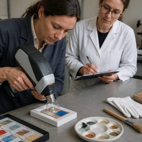
Using XRF for Non-Destructive Analysis
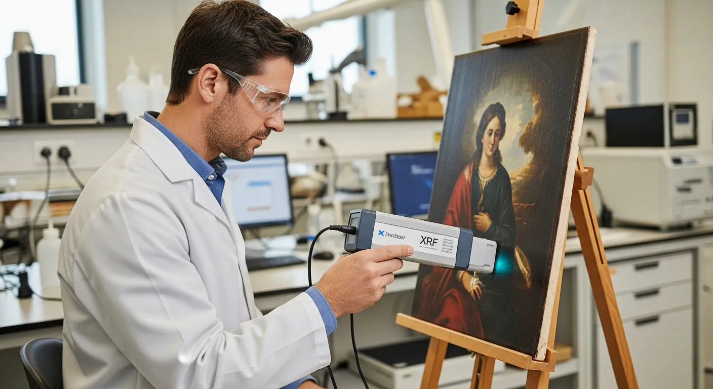
The non-destructive nature of X-ray Fluorescence Pigment Analysis makes it ideal for examining valuable artifacts. This technique allows for the analysis of an object without causing any physical damage.
This is especially important when studying historical objects. Preserving the integrity of the artifact is a primary concern.
Portable XRF instruments are available. These allow for in-situ analysis of artworks in museums or archaeological sites.
This eliminates the need to transport fragile objects to a laboratory. It reduces the risk of damage during handling.
- Identify pigments used in a painting
- Determine the elemental composition of ceramics
- Analyze the metallic components of sculptures
- Detect forgeries by identifying anachronistic pigments
- Map the distribution of elements in a sample
By using XRF, conservators can gain valuable information about the materials used in an artwork. This helps to inform conservation strategies.
It is possible to make informed decisions about cleaning, restoration, and storage. This ensures the long-term preservation of cultural heritage.
The non-destructive nature of XRF is particularly advantageous when dealing with objects that are considered national treasures or are of significant historical value. Traditional methods of analysis often require the removal of a sample, which can be unacceptable in these cases.
XRF allows for the analysis of the object without any physical alteration, preserving its integrity for future generations. This is crucial for maintaining the historical and cultural significance of the artifact.
The ability to perform in-situ analysis with portable XRF instruments further enhances the non-destructive aspect of the technique. Transporting fragile objects can be risky, and the process itself can cause damage.
Portable XRF allows conservators to bring the analysis to the object, eliminating the need for transportation and reducing the risk of damage. This is especially useful for large or immovable objects, such as murals or architectural elements.
The data obtained from XRF analysis can be used to create elemental maps of the object. These maps show the distribution of different elements across the surface of the object, providing valuable information about the materials used and their spatial relationships.
Elemental mapping can reveal hidden details about the object’s construction and history. It can also help to identify areas of deterioration or alteration.
XRF analysis can also be used to detect the presence of forgeries. By comparing the elemental composition of an object to known historical materials, it is possible to identify the use of anachronistic pigments or materials that were not available at the time the object was supposedly created.
This can be a powerful tool for art historians and collectors who are concerned about the authenticity of their objects. The non-destructive nature of XRF makes it an ideal technique for this type of analysis.
Case Studies: XRF in Pigment Authentication
XRF has been used in numerous case studies to authenticate artworks and identify forgeries. One notable example involves the analysis of Renaissance paintings.
XRF analysis revealed the presence of titanium white, a pigment not available until the 20th century, in a painting attributed to the 16th century. This discovery exposed the painting as a modern forgery.
In another case, XRF was used to examine a collection of ancient Chinese ceramics. The analysis identified the use of specific pigments and glaze compositions that were consistent with the purported period of origin.
This helped to confirm the authenticity of the ceramics. It provided valuable information about the techniques used by ancient artisans.
XRF has also been instrumental in studying the works of famous artists. By analyzing the pigments used by Van Gogh, researchers have gained insights into his artistic techniques and the materials he employed.
This type of analysis can help to differentiate between genuine works and imitations. It also sheds light on the artist’s creative process.
One compelling case study involved the examination of several paintings attributed to the Dutch master, Johannes Vermeer. XRF analysis was used to identify the pigments used in each painting, and the results were compared to known pigment palettes used by Vermeer and his contemporaries.
The analysis revealed that one of the paintings contained pigments that were not available during Vermeer’s lifetime. This strongly suggested that the painting was not authentic.
In another case, XRF was used to analyze a collection of ancient Egyptian artifacts. The analysis identified the presence of specific elements, such as copper and arsenic, which were characteristic of pigments used in ancient Egypt.
This helped to confirm the authenticity of the artifacts and provided valuable information about the materials and techniques used by ancient Egyptian artisans.
XRF has also been used to study the deterioration of pigments in historical artworks. By analyzing the elemental composition of degraded pigments, researchers can gain insights into the chemical processes that cause deterioration.
This information can be used to develop more effective conservation strategies to protect artworks from further damage. Understanding the chemical changes in pigments is crucial for long-term preservation.
The analysis of medieval manuscripts is another area where XRF has proven invaluable. The pigments used in illuminated manuscripts can provide information about the origin and date of the manuscript.
XRF can be used to identify these pigments without damaging the delicate pages of the manuscript. This allows researchers to study these important historical documents in a non-destructive manner.
In the investigation of potential art fraud, XRF serves as a critical tool for distinguishing genuine artifacts from imitations. The precise identification of pigments and materials can expose discrepancies that would otherwise remain undetected.
By analyzing the elemental composition, experts can determine if the materials used are consistent with the purported time period and origin of the artwork. This helps to ensure the integrity of the art market and protect collectors from fraudulent pieces.
Conclusion
X-ray fluorescence pigment analysis is a powerful tool for art conservation and art forensics. Its non-destructive nature, combined with its ability to provide detailed elemental information, makes it indispensable for studying historical pigments.
By using XRF, conservators and art historians can gain insights into artistic techniques. They can also authenticate artworks, and develop effective preservation strategies.
As technology advances, XRF instruments are becoming more portable and user-friendly. This will make the technique even more accessible to conservators and art historians around the world.
The continued development of XRF techniques promises to further enhance our understanding of historical pigments and artworks. This will contribute to the preservation of our cultural heritage for future generations.
The future of XRF in art conservation looks promising, with ongoing research focused on improving the sensitivity and spatial resolution of the technique. These advancements will enable even more detailed and accurate analyses of artworks.
By combining XRF with other analytical techniques, such as Raman spectroscopy and mass spectrometry, researchers can gain a more comprehensive understanding of the materials used in artworks. This multi-analytical approach is essential for addressing complex conservation challenges.
The use of XRF in art education is also becoming increasingly important. By teaching students about the principles and applications of XRF, we can ensure that the next generation of conservators and art historians are equipped with the skills and knowledge they need to protect our cultural heritage.
Ultimately, X-ray fluorescence pigment analysis plays a vital role in preserving and understanding our shared cultural heritage. Its ability to provide non-destructive, detailed elemental information makes it an invaluable tool for conservators, art historians, and other professionals working to protect and interpret our artistic legacy.