X-ray diffraction is a technique that can provide valuable insights into the composition and structure of materials. For art conservators and scientists, it offers a powerful, non-destructive method to identify the pigments used in historical artworks.
Understanding the chemical makeup of these pigments is essential for proper conservation and restoration efforts. This article will explore the principles of x-ray diffraction, its applications in pigment analysis, and its advantages and limitations.
By mastering this technique, we can better understand the materials used by artists throughout history. This knowledge allows for more informed decisions regarding the preservation of our cultural heritage.
The Fundamentals of X-Ray Diffraction
X-ray diffraction (XRD) is a technique used to determine the atomic and molecular structure of a crystalline material. It relies on the interaction of x-rays with the atoms in a sample.
When x-rays strike a crystalline material, they are scattered in various directions. The scattered x-rays can interfere with each other, either constructively or destructively, depending on the angles and spacing between the atoms.
Constructive interference occurs when the scattered waves are in phase, resulting in a strong signal. This phenomenon follows Bragg’s Law, which relates the angle of incidence of the x-rays to the spacing between the crystal lattice planes.
Bragg’s Law is expressed as nλ = 2dsinθ, where n is an integer, λ is the wavelength of the x-rays, d is the spacing between the lattice planes, and θ is the angle of incidence. By measuring the angles at which constructive interference occurs, the spacing between the lattice planes can be determined.
X-rays are electromagnetic radiation with wavelengths typically ranging from 0.01 to 10 nanometers. These wavelengths are comparable to the interatomic distances in crystalline materials, making them ideal for diffraction studies.

The interaction of x-rays with matter involves the excitation of electrons in the atoms of the sample. These excited electrons then re-emit x-rays, which are scattered in all directions.
The intensity of the diffracted x-rays is related to the arrangement of atoms in the crystal lattice. Therefore, by analyzing the diffraction pattern, we can gain insights into the crystal structure.
XRD is a powerful tool because it provides information about the long-range order in a material. This allows us to distinguish between crystalline and amorphous materials.
Crystalline materials exhibit sharp diffraction peaks due to the regular arrangement of atoms. Amorphous materials, on the other hand, produce broad, diffuse scattering patterns.
Sample Preparation for XRD Analysis
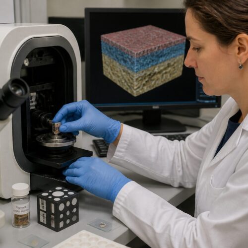
Proper sample preparation is crucial for obtaining accurate and reliable x-ray diffraction data. The goal is to present a representative sample to the x-ray beam in a way that minimizes errors.
For pigment analysis, samples are often in the form of small powder scrapings from a painting or artifact. These samples need to be carefully handled to avoid contamination or alteration.
The powder is then typically mounted onto a sample holder. One common method is to deposit the powder onto a glass slide or a zero-background holder, which minimizes the scattering from the holder itself.
Another approach involves mixing the powder with a binder, such as petroleum jelly or collodion, to form a paste. This paste is then applied to the sample holder, ensuring a smooth and even surface for analysis.
The particle size of the powder can also affect the quality of the diffraction pattern. Ideally, the powder should be finely ground to ensure that the x-rays interact with a representative number of crystallites.
If the particles are too large, the diffraction pattern may be spotty or uneven. This can lead to inaccuracies in the analysis.
In some cases, it may be necessary to use a mortar and pestle to grind the sample into a fine powder. However, care must be taken to avoid introducing contaminants during the grinding process.
The amount of sample required for XRD analysis is typically very small, often on the order of milligrams. This is advantageous for pigment analysis, where only limited amounts of sample are available.
The sample holder should be carefully cleaned before use to avoid contamination from previous samples. This is particularly important when analyzing trace amounts of pigments.
For certain applications, such as the analysis of thin films, specialized sample holders may be required. These holders are designed to ensure that the x-ray beam interacts with the sample at a specific angle.
Understanding Diffraction Patterns and Mineral Phases
The result of an x-ray diffraction experiment is a diffraction pattern, which is a plot of the intensity of the diffracted x-rays as a function of the diffraction angle (2θ). This pattern contains information about the crystalline phases present in the sample.
Each crystalline phase has a unique diffraction pattern, characterized by a set of peaks at specific angles. These peaks correspond to the different sets of lattice planes within the crystal structure.
| Mineral Phase | Chemical Formula | Key Diffraction Peaks (2θ) |
|---|---|---|
| Azurite | Cu3(CO3)2(OH)2 | 12.4°, 25.2°, 32.7° |
| Malachite | Cu2CO3(OH)2 | 24.9°, 34.2°, 36.0° |
| Hematite | Fe2O3 | 33.2°, 35.6°, 49.5° |
| Goethite | FeO(OH) | 21.3°, 36.6°, 55.2° |
The position of the diffraction peaks is determined by the size and shape of the unit cell of the crystal structure. The intensity of the peaks is related to the arrangement of atoms within the unit cell.
The width of the diffraction peaks can provide information about the crystallite size and the amount of strain in the crystal lattice. Broad peaks indicate small crystallite sizes or high levels of strain.
The presence of multiple crystalline phases in a sample will result in a diffraction pattern that is a superposition of the individual patterns of each phase. This can make the interpretation of the diffraction pattern more complex.
However, with careful analysis, it is possible to identify and quantify the different phases present in the sample. This is particularly useful in pigment analysis, where pigments may be mixtures of several components.
The identification of mineral phases is typically done by comparing the experimental diffraction pattern with reference patterns stored in databases. These databases contain diffraction patterns for a wide variety of crystalline materials.
The most widely used database is the Powder Diffraction File (PDF) maintained by the International Centre for Diffraction Data (ICDD). This database contains diffraction patterns for over 800,000 materials.
Software programs are available that can automatically search the database and identify the phases present in a sample. These programs use algorithms to match the experimental pattern with the reference patterns.
Identifying Pigments Based on Diffraction Data
The identification of pigments using x-ray diffraction relies on comparing the obtained diffraction pattern with reference patterns of known pigment materials. These reference patterns are typically stored in databases, such as the Powder Diffraction File (PDF) maintained by the International Centre for Diffraction Data (ICDD).
By matching the peak positions and intensities in the sample pattern with those in the reference patterns, the crystalline phases present in the pigment can be identified. This process often involves computer software that automates the pattern matching and phase identification.
For example, if the diffraction pattern shows peaks corresponding to the mineral azurite, it indicates the presence of the blue pigment azurite in the sample. Similarly, peaks corresponding to hematite would indicate the presence of the red pigment hematite.
The accuracy of pigment identification depends on the quality of the diffraction data and the completeness of the reference database. It’s important to consider that pigments may be mixed or altered over time, which can complicate the identification process.
Sometimes, pigments are present in very low concentrations, making their detection challenging. In such cases, advanced data processing techniques may be required to enhance the signal-to-noise ratio.
The presence of amorphous materials in the sample can also complicate the identification process. Amorphous materials do not produce sharp diffraction peaks, but rather a broad, diffuse scattering pattern.
This diffuse scattering can overlap with the peaks from crystalline pigments, making it difficult to accurately identify them. In some cases, it may be necessary to use other analytical techniques, such as Raman spectroscopy, to complement the XRD data.
The identification of pigments can also be complicated by the presence of alteration products. Over time, pigments can degrade or react with other materials, forming new crystalline phases.
These alteration products can produce their own diffraction peaks, which can interfere with the identification of the original pigments. It is important to be aware of the potential for alteration when interpreting diffraction patterns from historical pigments.
Quantitative Analysis using XRD
X-ray diffraction can also be used for quantitative analysis, which involves determining the relative amounts of different crystalline phases present in a sample. This is particularly useful in pigment analysis, where pigments may be mixtures of several components.
The most common method for quantitative analysis is the Rietveld refinement technique. This method involves fitting a calculated diffraction pattern to the experimental pattern, adjusting parameters such as the lattice parameters, atomic positions, and phase fractions to minimize the difference between the calculated and experimental patterns.
The Rietveld refinement provides an estimate of the weight fraction of each crystalline phase in the sample. This information can be used to determine the relative amounts of different pigments in a mixture.
However, quantitative analysis using x-ray diffraction can be challenging, especially when dealing with complex mixtures or poorly crystalline materials. The accuracy of the results depends on the quality of the diffraction data, the accuracy of the crystal structure models, and the proper accounting for factors such as preferred orientation and microabsorption.
Preferred orientation occurs when the crystallites in a sample are not randomly oriented. This can cause the intensities of certain diffraction peaks to be enhanced or suppressed.
Microabsorption occurs when the x-rays are absorbed differently by different phases in the sample. This can also affect the intensities of the diffraction peaks.
To obtain accurate quantitative results, it is important to correct for these effects. This can be done using various mathematical models and experimental techniques.
Another challenge in quantitative analysis is the presence of amorphous materials. Amorphous materials do not produce sharp diffraction peaks, making it difficult to quantify them using XRD.
In some cases, it may be possible to estimate the amount of amorphous material by measuring the background scattering in the diffraction pattern. However, this method is not very accurate.
Despite these challenges, quantitative analysis using XRD can provide valuable information about the composition of pigment mixtures. This information can be used to understand the artist’s techniques and the history of the artwork.
Advantages and Limitations of XRD in Pigment Analysis
X-ray diffraction offers several advantages for pigment analysis in cultural heritage conservation. It is a non-destructive technique, meaning that the sample is not significantly altered during the analysis.
XRD provides specific information about the crystalline phases present in a pigment, allowing for unambiguous identification of many common pigments. It can also be used to identify alteration products and degradation compounds, providing insights into the condition of the artwork.
- Non-destructive analysis
- Phase-specific identification
- Identification of alteration products
- Quantitative analysis potential
- Relatively simple sample preparation
However, XRD also has some limitations. It is only sensitive to crystalline materials. Amorphous materials, such as organic binders, cannot be directly identified using XRD.
The detection limit for minor components can be relatively high, meaning that trace amounts of pigments may not be detected. Sample preparation can be challenging, especially when dealing with very small or fragile samples.
The interpretation of diffraction patterns can be complex, especially when dealing with mixtures of pigments or altered materials. Expertise in crystallography and diffraction theory is required for accurate data analysis.
Another limitation is that XRD provides information about the bulk composition of the sample. It does not provide information about the spatial distribution of pigments within the artwork.
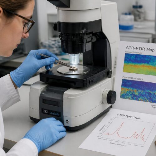
To overcome this limitation, it is often necessary to combine XRD with other analytical techniques, such as microscopy or spectroscopy. This allows for a more complete understanding of the composition and structure of the artwork.
Despite these limitations, XRD remains a valuable tool for pigment analysis in cultural heritage conservation. Its non-destructive nature and ability to provide phase-specific information make it an essential technique for studying historical artworks.
Case Studies: X-Ray Diffraction in Action
Several case studies demonstrate the effectiveness of x-ray diffraction in analyzing historical pigments. In one study, XRD was used to examine the pigments in a 15th-century Italian painting.
The analysis revealed the presence of azurite, vermilion, and lead white, providing insights into the artist’s palette. The identification of these pigments helped art historians understand the painting techniques used during that period.
Another case study involved the examination of pigments in ancient Egyptian artifacts. XRD analysis identified the presence of Egyptian blue, a synthetic pigment made from calcium copper silicate, in several artifacts.
This finding confirmed the use of this pigment in ancient Egypt and provided clues about the manufacturing techniques used to produce it. It’s fascinating how much information can be gleaned from tiny samples.
In a study of medieval illuminated manuscripts, XRD helped identify the use of gold particles in the illuminations. The presence of gold was confirmed by the characteristic diffraction peaks of metallic gold.
This finding provided insights into the materials and techniques used by medieval artists to create these beautiful works of art. It also helped to understand the economic value placed on these manuscripts.
XRD has also been used to study the degradation of pigments in historical artworks. In one study, XRD was used to identify the formation of lead carbonates on the surface of paintings containing lead white pigment.
This finding helped to understand the mechanisms of pigment degradation and to develop strategies for preventing further damage. Understanding these processes is key to preserving these artifacts for future generations.
These case studies illustrate the power of x-ray diffraction as a tool for studying historical pigments. By providing detailed information about the composition and structure of pigments, XRD can help us to understand the materials and techniques used by artists throughout history.
Advanced Techniques and Data Interpretation
While basic x-ray diffraction is a powerful tool, several advanced techniques can enhance its capabilities for pigment analysis. Micro-diffraction allows for the analysis of very small samples or specific areas of a painting.
This technique uses a focused x-ray beam to analyze areas as small as a few micrometers in diameter. Grazing incidence x-ray diffraction is used to analyze thin films or surface layers, providing information about the composition and structure of the surface.
Data interpretation is a critical aspect of x-ray diffraction analysis. It requires a thorough understanding of crystallography, diffraction theory, and the properties of pigment materials.
It’s important to consider factors such as peak broadening, preferred orientation, and the presence of amorphous materials when interpreting diffraction patterns. Careful data analysis and validation are essential for obtaining accurate and reliable results.
Synchrotron x-ray diffraction offers even greater capabilities due to the high intensity and tunable energy of synchrotron radiation. This allows for the analysis of even smaller samples and the detection of trace amounts of pigments.
Two-dimensional x-ray diffraction detectors can provide more detailed information about the orientation and distribution of crystallites in the sample. This can be particularly useful for studying the texture of pigments in paintings.
Rietveld refinement is a powerful technique for quantitative phase analysis, allowing for the accurate determination of the relative amounts of different pigments in a mixture. This requires careful modeling of the crystal structures of the pigments and the instrumental parameters.
The use of advanced software tools can greatly assist in data interpretation and analysis. These tools can automate tasks such as peak identification, phase identification, and quantitative analysis.
However, it is important to remember that these tools are only as good as the data that is input into them. Careful data collection and validation are essential for obtaining meaningful results.
Future Directions in X-Ray Diffraction Pigment Analysis
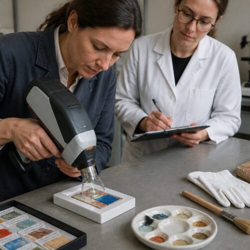
The field of x-ray diffraction pigment analysis is constantly evolving, with new techniques and applications emerging. One promising direction is the development of portable x-ray diffraction instruments.
These instruments allow for on-site analysis of artworks and artifacts, eliminating the need to transport samples to a laboratory. Another area of development is the integration of x-ray diffraction with other analytical techniques.
Combining x-ray diffraction with techniques such as Raman spectroscopy or x-ray fluorescence spectroscopy can provide a more complete understanding of the composition and structure of pigments. This multi-technique approach can provide complementary information, leading to more accurate and reliable pigment identification.
As technology advances, x-ray diffraction will continue to play a crucial role in the study and preservation of cultural heritage. These advancements will undoubtedly lead to new discoveries and insights into the materials and techniques used by artists throughout history.
The development of more sensitive detectors will allow for the analysis of even smaller samples and the detection of trace amounts of pigments. This will be particularly useful for studying degraded or altered pigments.
The use of machine learning and artificial intelligence can also help to improve the accuracy and efficiency of data interpretation. These techniques can be used to automatically identify patterns in diffraction data and to predict the composition of unknown pigments.
The integration of x-ray diffraction with three-dimensional imaging techniques, such as x-ray computed tomography, can provide a more complete understanding of the spatial distribution of pigments within artworks. This can help to reveal the layering and mixing of pigments used by artists.
The development of new reference databases and spectral libraries will also be crucial for improving the accuracy of pigment identification. These databases should include diffraction patterns for a wide variety of pigments, including both historical and modern materials.
By continuing to develop and refine x-ray diffraction techniques, we can gain a deeper understanding of the materials and techniques used by artists throughout history. This knowledge will help us to preserve our cultural heritage for future generations.
Conclusion
X-ray diffraction is an invaluable tool for the chemical analysis and conservation of historical pigments. Its ability to identify crystalline phases non-destructively makes it ideal for studying precious artworks and artifacts.
By understanding the principles of x-ray diffraction and its applications in pigment analysis, conservators and scientists can make informed decisions about the preservation of our cultural heritage. The insights gained from x-ray diffraction contribute to a deeper appreciation of the materials and techniques used by artists throughout history.
From identifying the components of ancient Egyptian blue to unraveling the secrets of Renaissance masterpieces, x-ray diffraction continues to be a cornerstone of art conservation science. Its impact on our understanding of art history is undeniable.
As analytical techniques continue to evolve, x-ray diffraction will remain a vital part of the conservator’s toolkit. This will ensure that future generations can experience and appreciate the artistic achievements of the past.
The ongoing development of portable instruments and advanced data analysis methods will further enhance the capabilities of x-ray diffraction. This will lead to new discoveries and a more complete understanding of the materials and techniques used by artists throughout history.
By combining x-ray diffraction with other analytical techniques, conservators and scientists can gain a holistic view of the composition, structure, and degradation of pigments. This multi-technique approach is essential for making informed decisions about the preservation of cultural heritage.
The knowledge gained from x-ray diffraction is not only valuable for conservation purposes, but also for art historical research. By understanding the materials and techniques used by artists, we can gain insights into their creative processes and the cultural context in which they worked.
X-ray diffraction has played a crucial role in authenticating artworks and identifying forgeries. By comparing the pigments used in a painting with those known to have been available during the artist’s lifetime, it is possible to detect inconsistencies that may indicate a forgery.
The future of x-ray diffraction in art conservation is bright. With ongoing advancements in technology and data analysis, this technique will continue to be an essential tool for preserving and understanding our cultural heritage.