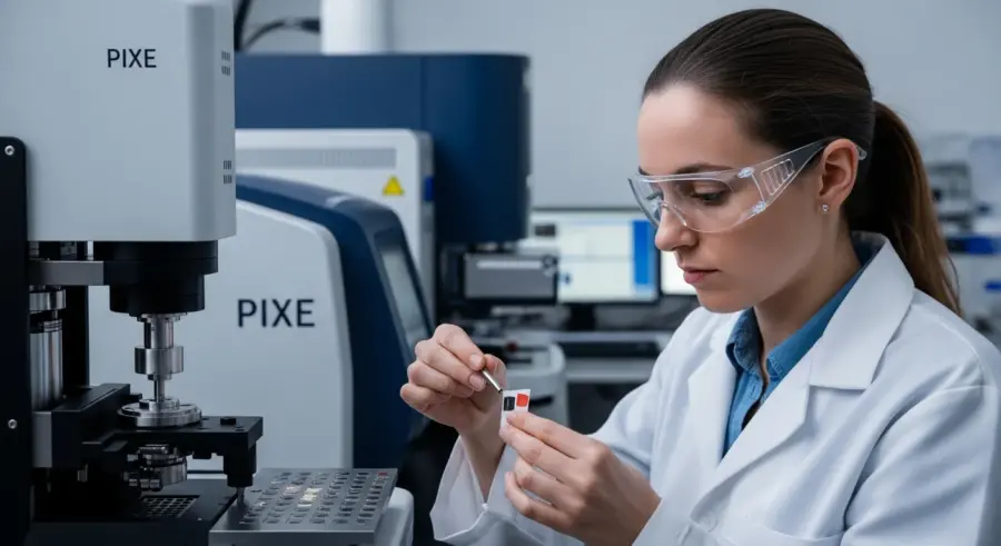
The world of art conservation is constantly evolving, with new technologies emerging to help us better understand and preserve our cultural heritage. One such technology is Particle-Induced X-ray Emission, or PIXE. This powerful analytical technique allows us to probe the elemental composition of materials in a non-destructive way, making it particularly valuable for the study of historical pigments.
By understanding the chemical makeup of pigments, conservators can gain insights into the artist’s techniques, the provenance of the materials, and the potential for degradation over time. PIXE analysis offers a unique window into the past, helping us to protect these precious artifacts for future generations.
This article will explore the principles behind particle induced x-ray emission pigments analysis, its applications in art conservation, and its advantages and limitations compared to other analytical techniques. We’ll also discuss the practical aspects of sample preparation and data interpretation, providing a comprehensive overview of this exciting field.
Understanding Particle-Induced X-ray Emission (PIXE)
Particle-Induced X-ray Emission, or PIXE, is a powerful analytical technique used to determine the elemental composition of a material. It works by bombarding a sample with a beam of energetic particles, typically protons or alpha particles, from a particle accelerator.
When these particles collide with the atoms in the sample, they can eject core electrons, creating vacancies in the inner electron shells. These vacancies are then filled by electrons from higher energy levels, resulting in the emission of X-rays with energies characteristic of the element involved.
By detecting and measuring the energy and intensity of these X-rays, we can identify the elements present in the sample and determine their concentrations. This process, known as ion beam analysis, provides a non-destructive way to analyze the elemental composition of a wide range of materials, including pigments.
The beauty of PIXE lies in its ability to provide quantitative elemental analysis with minimal sample preparation. It is a surface-sensitive technique, meaning it primarily analyzes the outer layers of the material, which is particularly useful for studying the composition of pigments in artworks.
The energetic particles used in PIXE are typically accelerated to energies in the MeV (Mega electron volt) range. This high energy is necessary to penetrate the electron cloud surrounding the atomic nucleus and interact with the core electrons.
The X-rays emitted during PIXE are not the only form of radiation produced. Other particles, such as backscattered ions, are also generated and can be used to obtain additional information about the sample.
The depth of analysis in PIXE depends on the energy of the incident particles and the composition of the sample. Typically, the analysis depth is on the order of a few micrometers, making it ideal for studying thin layers and surface coatings.
PIXE analysis is not limited to the study of pigments. It can also be used to analyze a wide range of other materials, including metals, ceramics, and polymers, making it a versatile tool for materials science and engineering.
The development of PIXE has revolutionized many fields, including archaeology, environmental science, and forensics. Its ability to provide rapid, non-destructive elemental analysis has made it an indispensable tool for scientists and researchers around the world.
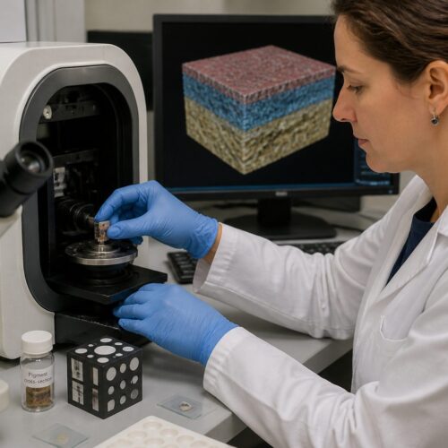
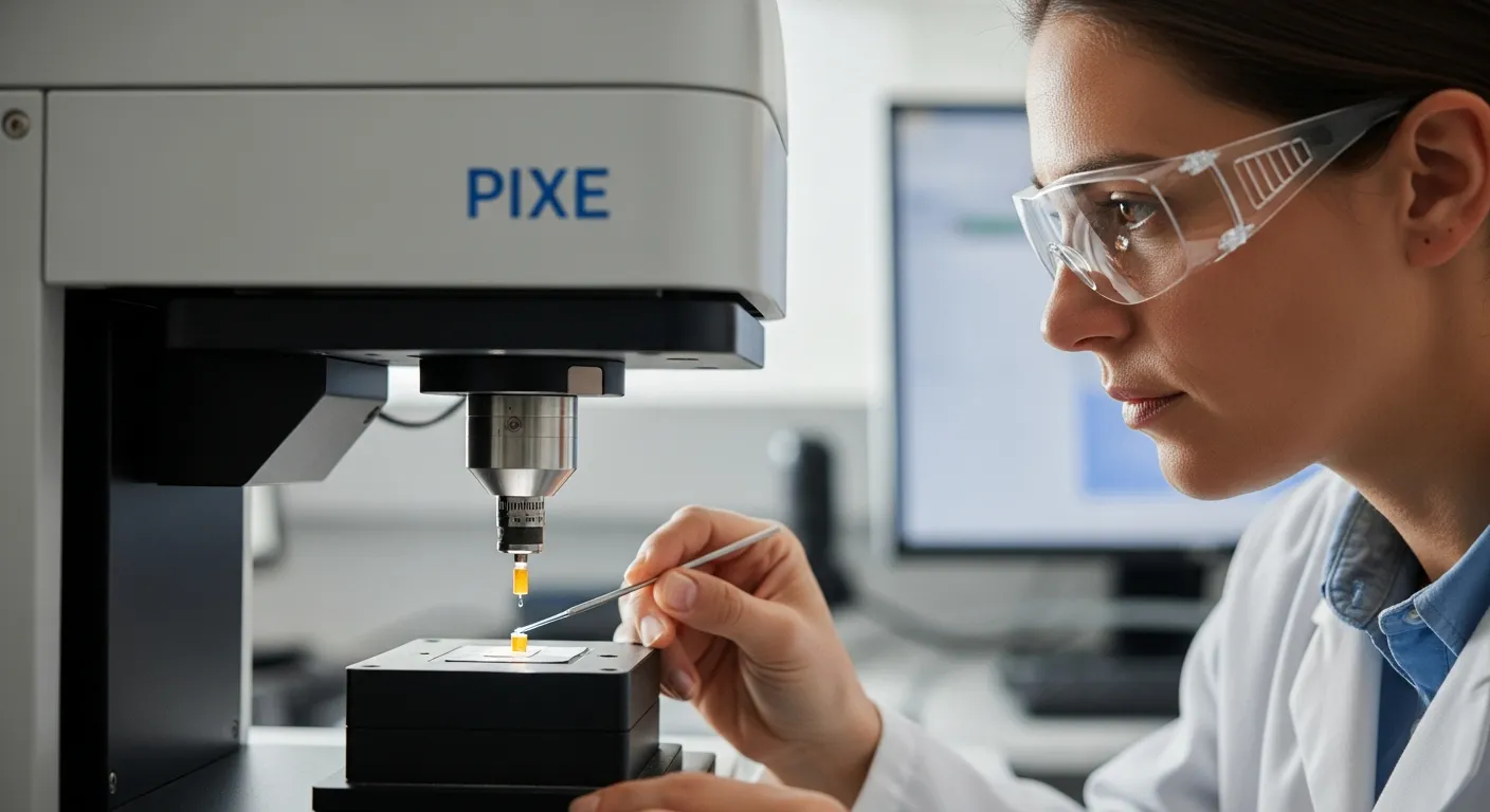
Sample Preparation for PIXE Analysis
Proper sample preparation is crucial for obtaining accurate and reliable results from PIXE analysis. While PIXE is often considered a non-destructive technique, some minimal intervention is usually required to prepare the sample for analysis.
For paintings and other artworks, this typically involves selecting a small area of interest and carefully cleaning the surface to remove any loose dirt or surface contaminants. In some cases, a tiny sample may be taken from an inconspicuous area of the artwork for separate analysis.
The sample is then mounted on a sample holder and placed in the path of the ion beam. It’s important to ensure that the sample is properly aligned and positioned to maximize the signal and minimize background noise.
For powdered pigments or small fragments, the sample can be mounted on a thin, clean substrate, such as carbon tape or a silicon wafer. Care must be taken to ensure that the sample is evenly distributed and that there are no gaps or voids that could affect the accuracy of the analysis.
The cleaning process is often performed using soft brushes, gentle solvents, or specialized cleaning solutions. The goal is to remove any surface debris without damaging the underlying pigment layers.
When taking a sample from an artwork, it is important to use a micro-sampling technique to minimize the impact on the object. This typically involves using a fine scalpel or a micro-drill to extract a tiny amount of material.
The choice of substrate for mounting powdered pigments depends on the elements being analyzed. Carbon tape is often used because it is relatively free of elemental contaminants, but other substrates may be more suitable for specific applications.
Proper grounding of the sample is essential to prevent charge buildup during PIXE analysis. Charge buildup can distort the ion beam and affect the accuracy of the results.
In some cases, it may be necessary to coat the sample with a thin layer of carbon or gold to improve its conductivity. This is particularly important for non-conductive materials such as ceramics and polymers.
Identifying Elements in Pigments Using PIXE
PIXE is particularly well-suited for identifying the elements present in historical pigments. Each element emits X-rays with characteristic energies, allowing for unambiguous identification.
For example, the presence of iron (Fe) in a pigment sample would indicate the use of iron oxides, such as ochre or sienna. Similarly, the detection of mercury (Hg) would suggest the presence of vermilion, a brilliant red pigment widely used in antiquity.
| Element | X-ray Energy (keV) | Associated Pigment |
|---|---|---|
| Iron (Fe) | 6.40 | Ochre, Sienna, Umber |
| Mercury (Hg) | 9.99 | Vermilion |
| Lead (Pb) | 10.55 | Lead White, Red Lead |
| Copper (Cu) | 8.04 | Verdigris, Azurite |
By comparing the measured X-ray energies with known values, we can create a detailed elemental profile of the pigment. This information can then be used to identify the specific pigments used in an artwork and to understand the artist’s palette.
The X-ray spectrum obtained from PIXE analysis is typically complex, with peaks corresponding to the different elements present in the sample. Deconvolution software is used to separate the overlapping peaks and identify the individual elements.
In addition to identifying the major elements in a pigment, PIXE can also detect trace elements. These trace elements can provide valuable information about the origin and processing of the pigment.
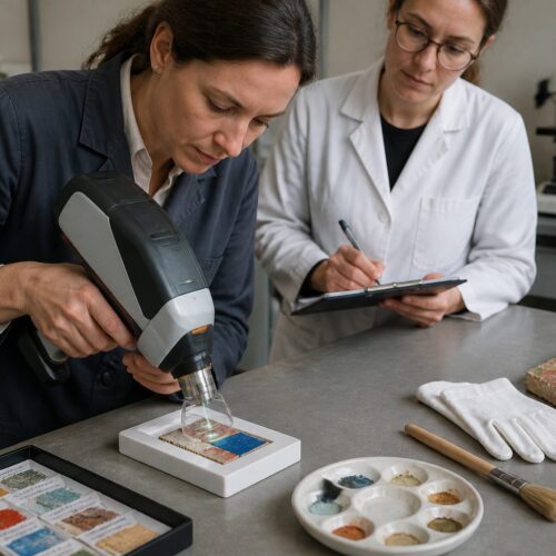
The identification of pigments using PIXE is often combined with other analytical techniques, such as Raman spectroscopy and X-ray diffraction, to provide a more complete characterization of the material.
The accuracy of pigment identification depends on the quality of the X-ray spectrum and the availability of reference data. A comprehensive database of X-ray energies and pigment compositions is essential for accurate analysis.
PIXE analysis can also be used to identify the presence of degradation products in pigments. These degradation products can provide information about the environmental conditions to which the artwork has been exposed.
Quantitative Analysis with PIXE
Beyond simply identifying the elements present, PIXE can also provide quantitative information about their concentrations. By carefully calibrating the instrument and measuring the intensity of the X-ray signals, we can determine the amount of each element present in the sample.
This quantitative analysis is crucial for understanding the composition of complex pigment mixtures and for detecting subtle variations in pigment recipes. For example, it can help us to distinguish between different grades of lead white or to identify the presence of extenders or adulterants in a pigment sample.
To perform quantitative analysis, the PIXE system must be calibrated using standards of known elemental composition. The measured X-ray intensities are then compared to the intensities from the standards to determine the concentrations of the elements in the unknown sample.
It’s important to note that quantitative PIXE analysis requires careful attention to detail and a thorough understanding of the matrix effects that can influence the X-ray intensities. These effects include absorption and fluorescence, which can alter the measured intensities and affect the accuracy of the results.
The accuracy of quantitative PIXE analysis depends on the quality of the standards used for calibration. The standards should be homogeneous, stable, and representative of the materials being analyzed.
Matrix effects can be corrected for using various mathematical models and algorithms. These corrections take into account the absorption and fluorescence of X-rays within the sample.
The detection limits for quantitative PIXE analysis depend on the element being analyzed and the composition of the sample. Typically, detection limits are in the parts per million (ppm) range.
Quantitative PIXE analysis can be used to determine the stoichiometry of pigments. This information can be used to identify the specific mineral or compound that is present in the sample.
The quantitative data obtained from PIXE analysis can be used to create elemental maps showing the distribution of different elements within the sample. These maps can provide valuable information about the layering and mixing of pigments.
Advantages and Limitations of PIXE in Pigment Analysis
PIXE offers several advantages over other analytical techniques for pigment analysis. Its non-destructive nature is a major benefit, allowing for the analysis of valuable artworks without causing significant damage.
PIXE is also a relatively fast and sensitive technique, capable of detecting trace elements in small samples. However, like any analytical method, PIXE has its limitations.
- Non-destructive or minimally destructive analysis
- High sensitivity for elemental detection
- Relatively fast analysis times
- Quantitative analysis capabilities
- Elemental mapping capabilities
One limitation is its surface sensitivity, which means that it primarily analyzes the outer layers of the material. This can be problematic if the surface is not representative of the bulk composition or if there are surface coatings or contaminants present.
Another limitation of PIXE is its inability to directly detect light elements, such as hydrogen, carbon, and oxygen. These elements are important components of many pigments, but they do not emit X-rays with sufficient energy to be detected by PIXE.
The cost of PIXE analysis can be a barrier for some researchers. PIXE facilities are typically located at large research institutions and require specialized equipment and trained personnel.
The interpretation of PIXE data can be complex and requires a thorough understanding of the underlying physics. Matrix effects and spectral interferences can complicate the analysis and require careful consideration.
Despite these limitations, PIXE remains a valuable tool for pigment analysis. Its advantages often outweigh its limitations, particularly when combined with other analytical techniques.
Compared to techniques like X-ray fluorescence (XRF), PIXE generally offers higher sensitivity and better spatial resolution. However, XRF instruments are often more readily available and less expensive to operate.
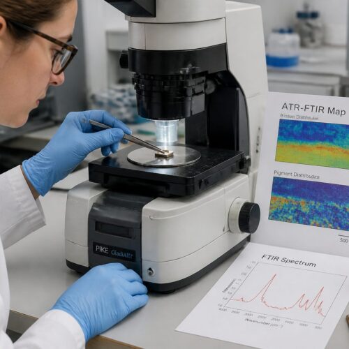
Elemental Mapping with PIXE
One of the most powerful features of PIXE is its ability to perform elemental mapping. By scanning the ion beam across the surface of a sample, we can create a map showing the distribution of different elements.
This is particularly useful for studying the layering and mixing of pigments in paintings. Elemental maps can reveal the presence of underlayers, retouching, or areas of pigment degradation.
To create an elemental map, the sample is rastered under the ion beam, and the X-ray intensities are recorded at each point. The resulting data is then processed to generate an image showing the distribution of each element.
The spatial resolution of the elemental map depends on the size of the ion beam and the step size used during scanning. By using a focused ion beam and small step sizes, it is possible to achieve high-resolution elemental maps that reveal fine details in the pigment distribution.
The color scale used to represent the elemental distribution in the map can be adjusted to highlight specific features. Different color palettes can be used to emphasize the presence of certain elements or to reveal subtle variations in concentration.
Elemental mapping can be used to identify areas of pigment mixing. By overlaying the maps for different elements, it is possible to see where pigments have been blended together by the artist.
The depth of analysis during elemental mapping can be controlled by adjusting the energy of the ion beam. This allows for the creation of three-dimensional elemental maps that show the distribution of elements at different depths within the sample.
Elemental mapping can be used to study the diffusion of elements within a pigment layer. This information can be used to understand the degradation mechanisms that are occurring within the artwork.
The data obtained from elemental mapping can be used to create quantitative maps showing the concentration of each element at each point on the sample. These quantitative maps can provide a more accurate representation of the pigment distribution than qualitative maps.
Applications of PIXE in Art Conservation
PIXE has found numerous applications in the field of art conservation. It has been used to study the pigments used in ancient Egyptian artifacts, Renaissance paintings, and modern artworks.
By identifying the pigments present, conservators can gain insights into the artist’s techniques and the materials available at the time. This information can be used to inform conservation treatments and to authenticate artworks.
For example, PIXE analysis has been used to study the degradation of pigments in paintings. By identifying the chemical changes that occur over time, conservators can develop strategies to slow down or prevent further degradation.
PIXE can also be used to study the effectiveness of conservation treatments. By analyzing the elemental composition of a treated area, conservators can determine whether the treatment has been successful in removing contaminants or stabilizing the pigments.
In the authentication of artworks, PIXE can help to determine whether the pigments used are consistent with the purported date of the artwork. The presence of pigments that were not available at the time the artwork was supposedly created can raise doubts about its authenticity.
PIXE can be used to study the composition of glazes and varnishes on artworks. This information can be used to understand the artist’s techniques and to develop appropriate cleaning and conservation strategies.
The analysis of ancient ceramics using PIXE can provide information about the provenance of the materials used to create the ceramics. This information can be used to trace trade routes and to understand the cultural exchange that occurred in ancient times.
PIXE can be used to study the corrosion of metals in archaeological artifacts. This information can be used to develop strategies to prevent further corrosion and to preserve these valuable objects for future generations.
The study of textiles using PIXE can provide information about the dyes used to color the textiles. This information can be used to understand the history of textile production and to develop appropriate conservation strategies.
Case Studies: PIXE in Action
Several case studies illustrate the power of PIXE in art conservation. One notable example is the study of the Vinča figurines, a collection of Neolithic artifacts from Serbia.
PIXE analysis revealed that the figurines were painted with a variety of pigments, including cinnabar (mercury sulfide) and hematite (iron oxide). The discovery of cinnabar, a relatively rare and expensive pigment, suggests that the Vinča culture had access to sophisticated trade networks.
Another case study involves the analysis of Renaissance paintings. PIXE was used to identify the pigments used by Renaissance artists, providing insights into their artistic techniques and the materials they had access to.
The analysis revealed that Renaissance artists used a wide range of pigments, including lead white, vermilion, azurite, and malachite. The presence of these pigments provides valuable information about the artistic practices of the time.
PIXE analysis was used to examine the Dead Sea Scrolls, revealing the composition of the inks used to write the ancient texts. The study helped to understand the writing practices and material availability during that historical period.
The analysis of Van Gogh’s paintings using PIXE has provided insights into the degradation of certain pigments, particularly the yellow chromates. This research helps conservators understand the changes occurring in the artworks and guides preservation efforts.
PIXE has been employed in the study of ancient Roman frescoes, helping to identify the pigments and techniques used by Roman artists. The findings contribute to our understanding of Roman art and technology.
Researchers have used PIXE to analyze the pigments in medieval illuminated manuscripts, providing valuable information about the materials and artistic practices of the time. The study contributes to the preservation and understanding of these precious cultural artifacts.
The use of PIXE in analyzing the Terracotta Army in China has revealed the composition of the pigments used to paint the statues. This information helps to reconstruct the original appearance of the army and understand the artistic techniques of the Qin Dynasty.
Future Directions in PIXE Analysis for Pigments
The field of PIXE analysis is constantly evolving, with new developments and applications emerging all the time. One area of active research is the development of portable PIXE systems.
These systems would allow for on-site analysis of artworks, eliminating the need to transport fragile objects to a laboratory. Another area of interest is the development of new data analysis techniques.
These techniques would allow for more accurate and detailed analysis of PIXE data, providing new insights into the composition and degradation of pigments. The future of PIXE analysis in art conservation looks bright.
As technology advances, PIXE will continue to play a crucial role in understanding and preserving our cultural heritage, particularly through improved elemental mapping and non-destructive analysis techniques.
The development of more sensitive detectors will allow for the analysis of even smaller samples and the detection of trace elements with greater accuracy. This will be particularly useful for studying highly degraded or fragile artworks.
The integration of PIXE with other analytical techniques, such as Raman spectroscopy and mass spectrometry, will provide a more comprehensive understanding of the composition and properties of pigments. This multi-technique approach will allow for the identification of organic and inorganic components, as well as the study of their interactions.
The use of artificial intelligence and machine learning algorithms will improve the efficiency and accuracy of PIXE data analysis. These algorithms can be trained to identify patterns in the data and to predict the composition and properties of pigments.
The development of new ion sources will allow for the use of different types of ions in PIXE analysis. This will expand the range of elements that can be detected and improve the sensitivity of the technique.
The application of PIXE to the study of modern and contemporary art will provide new insights into the materials and techniques used by artists in the 20th and 21st centuries. This will help conservators to develop appropriate strategies for the preservation of these artworks.
Conclusion
Particle-Induced X-ray Emission (PIXE) is a powerful tool for the chemical analysis and conservation of historical pigments. Its non-destructive nature, high sensitivity, and ability to perform elemental mapping make it an invaluable technique for studying artworks and artifacts.
By providing insights into the elemental composition of pigments, PIXE helps conservators understand the artist’s techniques, the provenance of materials, and the potential for degradation over time. As technology advances, PIXE will continue to play an increasingly important role in the field of art conservation, offering new ways to protect and preserve our cultural heritage.
From identifying pigments in ancient artifacts to monitoring the effectiveness of conservation treatments, PIXE has proven its worth as a valuable analytical tool. The continued development of portable PIXE systems and advanced data analysis techniques promises to further enhance its capabilities and expand its applications in the years to come.
With the ongoing research into ion beam analysis, we can expect even more refined methods for examining particle induced x-ray emission pigments, ensuring that future generations can appreciate the artistry and craftsmanship of the past. The study of particle induced x-ray emission pigments is a complex and ever-evolving field, but its importance in the world of art conservation cannot be overstated.
The combination of PIXE with other non-destructive techniques offers a comprehensive approach to art conservation, allowing for a deeper understanding of the materials and techniques used by artists throughout history. This collaborative approach ensures that our cultural heritage is preserved for future generations to enjoy and learn from.