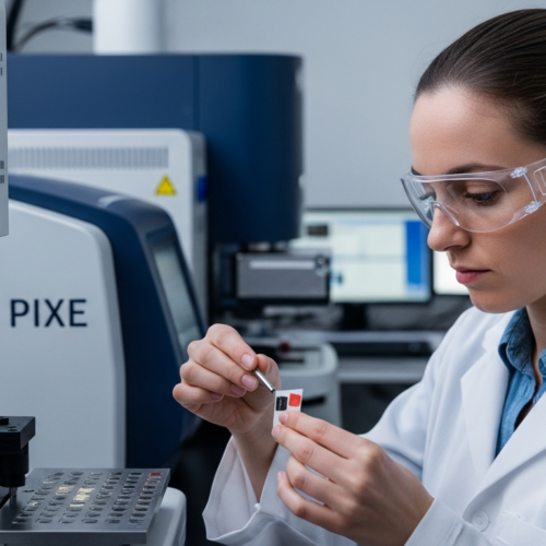
In the world of art conservation and historical research, understanding the composition of pigments is paramount. Knowing what makes up these colorful materials allows conservators to accurately restore artworks and scholars to gain insights into past artistic practices. Infrared spectroscopy pigment analysis, particularly using FTIR spectroscopy, has become an indispensable tool in this quest, providing detailed information about the molecular structure of pigments.
This technique offers a non-destructive means of identifying both organic and inorganic components, making it ideal for analyzing precious and fragile historical artifacts. By understanding the principles behind infrared spectroscopy pigment analysis, researchers and conservators can unlock a wealth of information hidden within the layers of paint.
This article will explore the basics of infrared spectroscopy, delve into sample preparation techniques, and discuss how to interpret FTIR spectra for pigment identification. We will also cover the advantages and limitations of this powerful analytical method, providing a comprehensive overview for anyone interested in the chemical analysis and conservation of historical pigments.
The Basics of Infrared Spectroscopy
Infrared spectroscopy, often referred to as FTIR spectroscopy (Fourier Transform Infrared spectroscopy), is a technique used to identify chemical compounds based on how they interact with infrared radiation. When a molecule absorbs infrared radiation, it vibrates at specific frequencies, which are determined by the types of bonds present and the masses of the atoms involved.
These vibrations include stretching, bending, scissoring, and rocking motions, each corresponding to a particular energy level. The absorption of infrared radiation at these specific frequencies creates a unique spectral fingerprint for each molecule, allowing for its identification and characterization.
At its core, infrared spectroscopy pigment analysis involves shining an infrared beam through a sample and measuring the amount of radiation that is transmitted or reflected. The resulting spectrum plots the intensity of the transmitted or reflected light as a function of wavenumber, which is inversely proportional to the wavelength of the infrared radiation.
Peaks in the spectrum correspond to specific vibrational modes within the molecule, and their positions and intensities provide information about the types of chemical bonds present. By comparing the spectrum of an unknown sample to reference spectra of known compounds, researchers can identify the components of the sample and determine its chemical composition.
The interaction between infrared radiation and a molecule is dictated by the molecule’s dipole moment. Only molecules with a change in dipole moment during vibration will absorb infrared radiation. This principle is crucial for understanding which vibrations will be active and contribute to the observed spectrum.

FTIR spectrometers use an interferometer to generate the infrared beam. This interferometer splits the beam into two paths, one fixed and one with a moving mirror, creating interference patterns. These patterns are then mathematically transformed using a Fourier transform to produce the final infrared spectrum.
The resolution of an FTIR spectrum is determined by the distance the moving mirror travels in the interferometer. Higher resolution spectra provide more detailed information about the vibrational modes of the molecule, but also require longer acquisition times.
Understanding the limitations of infrared spectroscopy is essential for accurate interpretation. Factors such as sample thickness, concentration, and temperature can all affect the appearance of the spectrum. Careful control of these variables is necessary for obtaining reliable results.
Sample Preparation Techniques for FTIR Analysis
Proper sample preparation is crucial for obtaining accurate and reliable results in infrared spectroscopy pigment analysis. The method used depends on the nature of the sample, whether it is a solid, liquid, or gas, and its physical properties.
For solid samples, such as pigment powders or paint chips, several techniques can be employed. One common method is the potassium bromide (KBr) pellet technique, where the sample is finely ground and mixed with KBr powder, which is transparent to infrared radiation.
The mixture is then pressed under high pressure to form a transparent pellet, which can be directly analyzed in the FTIR spectrometer. Another approach for solid samples is the use of attenuated total reflectance (ATR), which requires minimal sample preparation.
In ATR, the sample is placed in direct contact with an ATR crystal, such as diamond or germanium, and the infrared beam is passed through the crystal. The beam reflects off the interface between the crystal and the sample, and the resulting spectrum provides information about the sample’s surface composition. For liquid samples, a thin film can be applied to a salt plate (e.g., NaCl) and analyzed directly.
The KBr pellet technique requires careful grinding of the sample to ensure a homogeneous mixture. The particle size of the sample should be smaller than the wavelength of the infrared radiation to minimize scattering effects. It is also important to use high-quality KBr powder that is free from contaminants.
ATR spectroscopy is particularly useful for analyzing samples that are difficult to prepare as KBr pellets, such as hard or insoluble materials. The depth of penetration of the infrared beam into the sample is typically only a few micrometers, making ATR a surface-sensitive technique.
When using ATR, it is important to ensure good contact between the sample and the ATR crystal. This can be achieved by applying pressure to the sample using a pressure clamp. The choice of ATR crystal depends on the refractive index of the sample and the desired spectral range.
For liquid samples, it is important to use a solvent that is transparent in the infrared region of interest. Common solvents used in infrared spectroscopy include carbon tetrachloride, chloroform, and dichloromethane. The concentration of the sample should be optimized to obtain a good signal-to-noise ratio without saturating the detector.
Identifying Functional Groups and Pigment Components
Infrared spectroscopy pigment analysis excels at identifying the functional groups present in a sample, which are specific groups of atoms within a molecule that exhibit characteristic infrared absorption bands. These functional groups, such as carbonyls (C=O), hydroxyls (O-H), and amines (N-H), give rise to distinct peaks in the infrared spectrum, allowing researchers to identify the types of chemical bonds present in the sample.
By analyzing the positions and intensities of these peaks, it is possible to determine the presence of specific organic and inorganic components within a pigment. For example, the presence of a strong carbonyl peak around 1700 cm-1 indicates the presence of an ester or carboxylic acid, while a broad hydroxyl peak around 3300 cm-1 suggests the presence of alcohols or water.
| Functional Group | Bond | Approximate Wavenumber (cm-1) | Description |
|---|---|---|---|
| Alkanes | C-H | 2850-2960 | Strong, sharp peaks |
| Alkenes | C=C | 1640-1680 | Medium intensity |
| Alcohols | O-H | 3200-3600 | Broad peak |
| Carbonyls | C=O | 1650-1800 | Strong, sharp peak |
| Amines | N-H | 3300-3500 | Medium intensity, one or two peaks |
The position of a functional group peak can be influenced by its chemical environment. Factors such as hydrogen bonding, inductive effects, and resonance can shift the peak position to higher or lower wavenumbers. Careful consideration of these effects is necessary for accurate identification of functional groups.
The intensity of a functional group peak is related to the concentration of the functional group in the sample and the strength of its dipole moment. Stronger dipole moments result in more intense peaks. The intensity of a peak can be used to estimate the relative abundance of a functional group in the sample.
The shape of a functional group peak can also provide valuable information. Broad peaks are often associated with hydrogen bonding, while sharp peaks are typically associated with isolated functional groups. The shape of a peak can be used to distinguish between different types of functional groups.
The fingerprint region of the infrared spectrum, which is typically between 1500 and 500 cm-1, contains complex patterns of peaks that are unique to each molecule. This region is often used to identify specific compounds by comparing the spectrum to reference spectra. The fingerprint region can be particularly useful for distinguishing between isomers or closely related compounds.
Analyzing Organic and Inorganic Pigments with FTIR
Infrared spectroscopy pigment analysis is a versatile technique that can be used to analyze both organic and inorganic pigments. Organic pigments, which are typically derived from natural sources or synthesized from organic compounds, exhibit complex infrared spectra due to their intricate molecular structures.
These spectra often contain numerous peaks corresponding to various functional groups, such as aromatic rings, carbonyls, and amines, providing a wealth of information about the pigment’s chemical composition. For example, the infrared spectrum of indigo, a natural organic pigment, shows characteristic peaks associated with its indolic structure, including C=O stretching vibrations and N-H bending vibrations.
Similarly, the spectra of synthetic organic pigments, such as phthalocyanine blue and quinacridone red, exhibit distinct peaks related to their complex aromatic and heterocyclic structures. In contrast, inorganic pigments, which are typically metal oxides, sulfides, or carbonates, tend to have simpler infrared spectra with fewer, broader peaks.
These peaks are primarily associated with the vibrational modes of the metal-oxygen or metal-sulfur bonds within the pigment’s crystal lattice. For instance, the infrared spectrum of hematite (iron oxide) shows strong absorption bands due to Fe-O stretching vibrations, while the spectrum of lead white (lead carbonate) exhibits peaks related to Pb-O and C-O stretching vibrations.
The analysis of organic pigments often involves identifying the specific chromophore responsible for the pigment’s color. The chromophore is the part of the molecule that absorbs light in the visible region of the spectrum. The infrared spectrum can provide information about the structure and bonding of the chromophore.
The analysis of inorganic pigments often involves identifying the specific metal cation and anion present in the pigment. The infrared spectrum can provide information about the vibrational modes of the metal-oxygen or metal-sulfur bonds, which are characteristic of the specific metal cation and anion.
The presence of impurities or additives in the pigment can also affect the infrared spectrum. These impurities or additives may give rise to additional peaks in the spectrum or alter the positions and intensities of the existing peaks. Careful analysis of the spectrum is necessary to identify and account for the presence of impurities or additives.
The particle size and morphology of the pigment can also affect the infrared spectrum. Smaller particle sizes and irregular morphologies can lead to increased scattering of the infrared radiation, which can affect the intensity and shape of the peaks. The sample preparation technique should be optimized to minimize these effects.
Interpreting FTIR Spectra for Pigment Identification
Interpreting FTIR spectra for pigment identification requires a systematic approach that involves comparing the spectrum of an unknown sample to reference spectra of known pigments. This process typically begins with identifying the major peaks in the spectrum and assigning them to specific functional groups or vibrational modes.
Reference databases, such as those maintained by universities and conservation institutions, are invaluable resources for this task, providing spectra of a wide range of pigments and other materials commonly found in artworks. By comparing the peak positions and intensities in the unknown spectrum to those in the reference spectra, it is often possible to narrow down the list of possible pigments and identify the components of the sample.
However, it is important to note that pigment mixtures can complicate the interpretation process, as the spectrum may contain peaks from multiple pigments. In such cases, spectral deconvolution techniques can be used to separate the contributions from each individual pigment and facilitate identification.
Additionally, factors such as pigment degradation and the presence of binding media can also affect the infrared spectrum, making interpretation more challenging. Careful consideration of these factors and the use of complementary analytical techniques, such as Raman spectroscopy and X-ray diffraction, can help to improve the accuracy and reliability of pigment identification.
The first step in interpreting an FTIR spectrum is to normalize the spectrum to a common baseline. This removes any background absorption or scattering effects that may be present in the spectrum. Normalization allows for a more accurate comparison of the peak intensities.
The next step is to identify the major peaks in the spectrum and assign them to specific functional groups or vibrational modes. This can be done by consulting reference tables or databases that list the characteristic infrared absorption bands for various functional groups and compounds. It’s important to consider the shape and intensity of the peaks as well as their position.
Once the major peaks have been assigned, the spectrum can be compared to reference spectra of known pigments. This can be done visually or by using spectral matching algorithms. It is important to consider the possibility of pigment mixtures and to look for peaks that are characteristic of multiple pigments.
Finally, the results of the FTIR analysis should be compared to the historical context of the artwork or artifact. This can help to confirm the identification of the pigments and to provide insights into the artist’s techniques and materials. The geographical origin and time period of the artwork can provide valuable clues.
Advantages and Limitations of FTIR in Pigment Analysis
Infrared spectroscopy pigment analysis offers several advantages that make it a valuable tool for art conservation and historical research. One of the most significant advantages is its non-destructive nature, particularly when using techniques like ATR, which allows for the analysis of precious and fragile artifacts without causing damage.
FTIR spectroscopy also provides detailed information about the molecular structure of pigments, allowing for the identification of both organic and inorganic components. Despite its strengths, infrared spectroscopy pigment analysis also has some limitations that must be considered.
- Sensitivity can be limited for minor components
- Spectral interpretation can be complex for mixtures
- Water interference can obscure certain regions
- Sample preparation can be time-consuming
- Reference spectra may not always be available
The sensitivity of FTIR spectroscopy can be limited by the concentration of the pigment in the sample and the strength of its infrared absorption. Minor components may not be detectable if they are present at low concentrations or if their infrared absorption is weak. Signal enhancement techniques, such as spectral averaging, can be used to improve the sensitivity of the analysis.
Spectral interpretation can be challenging for complex mixtures of pigments, as the peaks from multiple pigments may overlap. Spectral deconvolution techniques can be used to separate the contributions from each individual pigment, but these techniques require careful optimization and may not always be successful. Expert knowledge is often required for accurate interpretation.
Water interference can be a significant problem in FTIR spectroscopy, as water absorbs strongly in the infrared region. Water vapor in the air can also contribute to the background spectrum. It is important to dry the sample thoroughly before analysis and to purge the spectrometer with dry air or nitrogen to minimize water interference.
Sample preparation can be time-consuming, especially for solid samples that require grinding and mixing with KBr. The sample preparation technique must be carefully optimized to ensure that the sample is homogeneous and free from contaminants. Automation of sample preparation can help to reduce the time and effort required.
Case Studies: Practical Applications of FTIR
To better illustrate the power of infrared spectroscopy pigment analysis, let’s consider a few case studies where this technique has provided valuable insights. In one study, FTIR spectroscopy was used to analyze the pigments in a 15th-century Italian painting.
The analysis revealed the presence of azurite, vermilion, and lead white, which were common pigments used during that period. This information helped art historians to understand the artist’s palette and techniques, as well as to authenticate the painting.
In another case, FTIR spectroscopy was employed to investigate the degradation of pigments in a set of ancient Egyptian artifacts. The analysis showed that the blue pigments, which were originally vibrant, had degraded over time due to the formation of copper chlorides and other degradation products.
This finding helped conservators to develop appropriate conservation strategies to stabilize the pigments and prevent further deterioration. In a third example, infrared spectroscopy pigment analysis was used to identify the pigments used in a series of historical documents.
The documents were suspected of being forgeries, and the pigment analysis was used to determine whether the pigments were consistent with the time period in which the documents were supposedly created. The analysis revealed that some of the pigments were not available until centuries later, confirming that the documents were indeed forgeries. This demonstrates the use of FTIR in forensic science.
In the conservation of a Dutch Golden Age painting, FTIR was crucial in identifying a discolored varnish layer. Knowing the chemical composition of the varnish allowed conservators to select a suitable solvent for its removal without damaging the underlying paint layers. This targeted approach minimized the risk of unintended consequences.
FTIR has also been employed to analyze the pigments used in prehistoric cave paintings. By identifying the pigments, researchers can gain insights into the materials available to early humans and their artistic practices. This provides a window into the past, revealing details about ancient cultures.
In the study of illuminated manuscripts, FTIR can help to identify the pigments used in the intricate decorations. This information can be used to determine the origin and date of the manuscript, as well as to understand the artistic techniques employed by the illuminator. It contributes to a deeper understanding of the manuscript’s history and significance.
Advanced Techniques and Future Directions
While conventional FTIR spectroscopy is a powerful tool, several advanced techniques have been developed to enhance its capabilities and address some of its limitations. One such technique is infrared microscopy, which allows for the analysis of very small samples or specific areas within a larger sample.
Infrared microscopy can be used to identify pigments in individual paint layers or to analyze microscopic inclusions within a pigment particle. Another advanced technique is two-dimensional correlation spectroscopy (2D-COS), which enhances the spectral resolution and sensitivity of FTIR spectroscopy by correlating variations in the spectrum as a function of an external perturbation, such as temperature or pressure.
2D-COS can be used to resolve overlapping peaks and identify subtle spectral features that would be difficult to detect with conventional FTIR spectroscopy. Looking ahead, future directions in infrared spectroscopy pigment analysis include the development of more sensitive and portable instruments, as well as the creation of more comprehensive reference databases.
The integration of machine learning and artificial intelligence algorithms into the spectral interpretation process also holds great promise for improving the accuracy and efficiency of pigment identification. These advancements will undoubtedly further enhance the role of infrared spectroscopy pigment analysis in art conservation and historical research.
Synchrotron-based FTIR microspectroscopy offers even higher spatial resolution and sensitivity, allowing for the analysis of even smaller samples and more dilute pigments. This technique is particularly useful for analyzing layered structures and identifying trace amounts of pigments.
Combining FTIR with other analytical techniques, such as Raman spectroscopy and X-ray diffraction, can provide a more comprehensive understanding of the pigment’s composition and structure. This multi-technique approach can help to overcome the limitations of each individual technique and provide more accurate and reliable results.
The development of portable FTIR instruments is allowing researchers to perform analyses in situ, without having to remove samples from the artwork or artifact. This is particularly useful for analyzing large or fragile objects that cannot be easily transported to a laboratory.
The use of spectral libraries and databases is becoming increasingly important for pigment identification. These libraries contain reference spectra for a wide range of pigments and other materials, allowing researchers to quickly and easily compare their spectra to known standards. Standardized data formats and open-source databases are facilitating the sharing of spectral information.
Ensuring Accuracy and Reliability in FTIR Analysis
To ensure the accuracy and reliability of infrared spectroscopy pigment analysis, it is essential to follow established protocols and best practices. Proper calibration of the FTIR spectrometer is crucial to ensure that the instrument is accurately measuring the infrared spectrum.
This involves running standard reference materials and adjusting the instrument parameters to match the known spectrum of the reference material. Careful sample preparation is also essential to minimize errors and artifacts in the spectrum.
This includes ensuring that the sample is homogeneous, free from contaminants, and properly mounted in the spectrometer. When interpreting the spectrum, it is important to consider the limitations of the technique and to use multiple lines of evidence to support the identification of pigments.
This may involve comparing the FTIR spectrum to reference spectra, consulting with experts in the field, and using complementary analytical techniques to confirm the results. By following these guidelines, researchers and conservators can ensure that their infrared spectroscopy pigment analysis is accurate, reliable, and meaningful.
Regularly check the performance of the FTIR instrument using certified reference materials. This helps to identify any drift or degradation in the instrument’s performance. Consistent monitoring ensures data integrity over time.
Implement strict quality control measures for sample preparation. This includes using clean glassware, high-purity solvents, and appropriate handling techniques. Minimizing contamination is crucial for accurate results.
Use appropriate spectral processing techniques, such as baseline correction, smoothing, and atmospheric compensation. These techniques can help to improve the quality of the spectrum and reduce noise. Proper data processing enhances the clarity of the results.
Document all aspects of the analysis, including the sample preparation method, instrument parameters, and spectral interpretation. This ensures traceability and allows for independent verification of the results. Thorough documentation is essential for scientific rigor.
Conclusion
Infrared spectroscopy pigment analysis, particularly FTIR spectroscopy, stands as a powerful and versatile technique for understanding the composition of historical pigments. Its non-destructive nature, combined with its ability to identify both organic and inorganic components, makes it an indispensable tool for art conservation and historical research.
By understanding the principles behind infrared spectroscopy pigment analysis, employing proper sample preparation techniques, and carefully interpreting the resulting spectra, researchers and conservators can unlock a wealth of information hidden within the layers of paint.
As technology continues to advance, the capabilities of infrared spectroscopy pigment analysis will only continue to grow, further enhancing its role in preserving and understanding our cultural heritage. The future of pigment analysis looks brighter than ever, thanks to these incredible scientific advancements.
The ongoing development of more sophisticated software and algorithms will further streamline the process of spectral interpretation. This will enable researchers to analyze complex datasets more efficiently and accurately. Automation and artificial intelligence will play an increasingly important role.
The increasing availability of open-access spectral libraries will facilitate collaboration and data sharing among researchers worldwide. This will lead to a more comprehensive understanding of pigment composition and degradation processes. Open science initiatives will drive further progress.
The integration of FTIR with other analytical techniques will provide a more holistic view of the materials used in art and historical artifacts. This multi-faceted approach will lead to new discoveries and insights. Interdisciplinary research will be key to unlocking the full potential of these techniques.
Ultimately, infrared spectroscopy pigment analysis will continue to play a vital role in preserving our cultural heritage for future generations. By understanding the materials used in the past, we can better protect and appreciate the art and artifacts that tell the story of human civilization. This is a critical mission that requires ongoing innovation and collaboration.