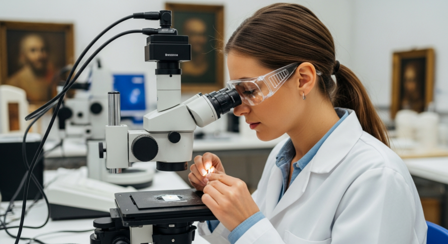
The world of art conservation is a fascinating blend of science and art. Preserving cultural heritage requires a deep understanding of the materials used by artists throughout history. One of the most powerful tools in a conservator’s arsenal is microscopy, allowing for detailed examination of pigment composition and structure.
Microscopy pigment analysis provides invaluable insights into the authenticity, age, and condition of artworks. This guide will explore the various microscopy techniques used in pigment analysis, offering a comprehensive overview for art enthusiasts and conservation professionals alike.
From polarized light microscopy to scanning electron microscopy and Raman microscopy, each method offers unique advantages in identifying and characterizing pigments. Understanding these techniques is essential for unlocking the secrets hidden within historical artworks and ensuring their preservation for future generations.
Understanding the Basics of Microscopy
Microscopy is the science of viewing small objects and structures that are not visible to the naked eye. It involves using microscopes to magnify these objects, revealing details about their morphology, composition, and arrangement. This magnification allows researchers and conservators to study materials at a level of detail previously unattainable.
In the context of pigment analysis, microscopy enables the identification of individual pigment particles. It also helps in understanding how these particles interact with each other and the surrounding binding medium. Different types of microscopy offer varying levels of magnification and analytical capabilities, making them suitable for diverse research questions.
Optical microscopy, also known as light microscopy, is one of the most common and accessible techniques. It uses visible light and a system of lenses to magnify the image of a sample. The magnification range typically extends up to 1000x, providing sufficient detail for identifying many common pigments.
Electron microscopy, on the other hand, uses a beam of electrons to illuminate the sample. This technique offers significantly higher magnification, up to millions of times, allowing for the visualization of extremely fine details. Electron microscopy is particularly useful for studying the morphology of pigment particles and identifying trace elements within them.
Preparing Pigment Samples for Microscopic Analysis
Proper sample preparation is crucial for obtaining accurate and reliable results from microscopy pigment analysis. The methods used to prepare samples depend on the type of microscopy being employed and the nature of the artwork being studied. Minimizing damage to the artwork during sampling is of utmost importance.

For optical microscopy, samples are often prepared as small cross-sections or dispersed particles on a glass slide. Cross-sections involve taking a tiny sample of the paint layer and embedding it in a resin. This allows for the examination of the layer structure and the distribution of pigments within each layer.
Dispersed particle mounts involve gently scraping a small amount of pigment from the artwork and suspending it in a liquid medium on a glass slide. The particles are then spread out to allow for individual examination. This method is particularly useful for identifying pigments based on their color, shape, and optical properties.
For electron microscopy, samples typically need to be coated with a thin layer of conductive material, such as gold or carbon. This coating helps to prevent the buildup of static charge on the sample, which can distort the image. Sample preparation for electron microscopy requires specialized equipment and expertise to ensure that the sample is not damaged during the process.
Polarized Light Microscopy: Identifying Crystalline Structures
Polarized light microscopy (PLM) is a powerful technique for identifying crystalline materials, including many historical pigments. PLM utilizes the properties of polarized light to reveal the optical characteristics of these materials. This technique is particularly valuable for distinguishing between different types of pigments that may appear similar under normal light.
When polarized light passes through a crystalline material, it is split into two rays that travel at different speeds. This phenomenon, known as birefringence, causes the light to emerge with different polarization states. The resulting interference patterns can be observed using an analyzer, providing information about the crystal structure and orientation of the material.
| Pigment | Color | Birefringence |
|---|---|---|
| Azurite | Blue | Strong |
| Malachite | Green | Moderate |
| Vermilion | Red | Very High |
| Ultramarine | Blue | Low |
Scanning Electron Microscopy (SEM): Visualizing Pigment Morphology
Scanning electron microscopy (SEM) is a technique that uses a focused beam of electrons to create high-resolution images of a sample’s surface. SEM is particularly useful for visualizing the morphology, or shape and texture, of pigment particles. The high magnification and depth of field offered by SEM make it an invaluable tool for pigment analysis.
In SEM, the electron beam scans across the surface of the sample, and the emitted secondary electrons are detected to create an image. The intensity of the secondary electron signal depends on the angle of the sample’s surface relative to the beam. This creates a three-dimensional-like image that reveals fine details of the pigment particles.
SEM can reveal important information about the size, shape, and aggregation state of pigment particles. For example, some pigments consist of well-defined, crystalline particles, while others are amorphous or aggregated. This information can be used to differentiate between pigments and to assess the quality of the pigment material.
Furthermore, SEM can be used to examine the surface of pigment particles for signs of degradation or alteration. Changes in surface texture or the presence of corrosion products can indicate the age and condition of the pigment. This information is crucial for understanding the long-term stability of pigments in artworks.
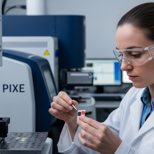
Energy-Dispersive X-ray Spectroscopy (EDS): Elemental Composition Analysis
Energy-dispersive X-ray spectroscopy (EDS) is an analytical technique used in conjunction with SEM to determine the elemental composition of a sample. When the electron beam in SEM interacts with the sample, it causes the emission of characteristic X-rays. These X-rays have energies that are specific to the elements present in the sample.
EDS detects and measures the energy of these X-rays, providing a quantitative analysis of the elemental composition of the pigment particles. This information is essential for identifying the chemical compounds that make up the pigment. By knowing the elemental composition, conservators can accurately identify the pigment and learn more about its origin and manufacturing process.
For example, EDS can differentiate between different types of blue pigments, such as azurite (copper carbonate) and ultramarine (sodium aluminum silicate with sulfur). Azurite will show the presence of copper, carbon, and oxygen, while ultramarine will show the presence of sodium, aluminum, silicon, oxygen, and sulfur. This level of specificity is invaluable for accurate pigment identification.
EDS can also be used to detect trace elements within pigment particles. These trace elements can provide clues about the source of the pigment material. For instance, the presence of certain impurities in lead white pigment can indicate the geographical origin of the lead ore used to make the pigment. This information can be useful for provenance studies and for understanding trade routes.
Raman Microscopy: Molecular Vibrational Analysis
Raman microscopy is a spectroscopic technique that provides information about the molecular vibrations within a sample. When a laser beam is focused on a sample, some of the light is scattered inelastically, resulting in a shift in wavelength. This shift, known as the Raman effect, is specific to the vibrational modes of the molecules present in the sample.
By analyzing the Raman spectrum, which is a plot of the intensity of the scattered light versus the Raman shift, it is possible to identify the molecular composition of the pigment. Raman microscopy is particularly useful for identifying organic pigments, which may be difficult to identify using other techniques. It can also be used to differentiate between different crystalline forms of the same pigment.
Raman microscopy is a non-destructive technique, meaning that it does not damage the sample during analysis. This is a significant advantage when studying valuable artworks. The technique can be used to analyze pigments in situ, without the need for sample preparation. This minimizes the risk of altering or contaminating the sample.
Furthermore, Raman microscopy can provide information about the degradation products of pigments. As pigments age, they may undergo chemical changes that alter their molecular structure. These changes can be detected by Raman microscopy, providing insights into the condition and stability of the pigment.
Interpreting Microscopic Data for Pigment Identification
Interpreting microscopic data for pigment identification requires a combination of expertise, experience, and reference materials. Each microscopy technique provides a different type of information, and it is important to integrate these data to arrive at an accurate identification. Reference databases and spectral libraries are essential tools for this process.
For polarized light microscopy, the birefringence, color, and morphology of the pigment particles are compared to known reference materials. Atlases of polarized light micrographs of common pigments are available to aid in this process. These atlases provide detailed descriptions and images of the optical properties of various pigments.
For scanning electron microscopy and energy-dispersive X-ray spectroscopy, the morphology and elemental composition of the pigment particles are compared to reference data. Databases of elemental compositions for common pigments are available. These databases allow conservators to match the elemental signature of an unknown pigment to a known reference.
For Raman microscopy, the Raman spectrum of the pigment is compared to spectral libraries of known pigments. These libraries contain Raman spectra for a wide range of organic and inorganic pigments. Spectral matching algorithms can be used to identify the pigment based on its Raman spectrum. Careful interpretation is needed, as mixtures of pigments can produce complex spectra.
Case Studies: Microscopy in Action
Microscopy pigment analysis has played a crucial role in numerous art conservation projects. These case studies demonstrate the power of microscopy in solving complex problems related to authenticity, dating, and conservation. From identifying rare pigments to uncovering forgeries, microscopy has proven to be an invaluable tool.
One example is the analysis of paintings attributed to Vincent van Gogh. Microscopy has been used to identify the pigments used by Van Gogh and to compare them to those used by other artists of the period. This has helped to authenticate paintings and to understand Van Gogh’s artistic techniques.
Another case study involves the examination of medieval illuminated manuscripts. Microscopy has been used to identify the pigments used to create the vibrant colors in these manuscripts. This information has provided insights into the materials available to medieval artists and the techniques they used to create their masterpieces.
Microscopy has also been used to study the degradation of pigments in artworks. By examining the surface of pigment particles, conservators can identify signs of chemical alteration and corrosion. This information can be used to develop conservation strategies to prevent further damage to the artwork.
The Future of Microscopy in Art Conservation
The field of microscopy is constantly evolving, with new techniques and technologies being developed all the time. These advances promise to further enhance the capabilities of microscopy pigment analysis in art conservation. Improved resolution, sensitivity, and automation are driving the future of this field.
One promising area of development is the use of super-resolution microscopy. These techniques can overcome the diffraction limit of light, allowing for even finer details to be visualized. Super-resolution microscopy could reveal new information about the structure and composition of pigments at the nanoscale.
Another area of development is the integration of microscopy with other analytical techniques. Combining microscopy with mass spectrometry, for example, could provide even more detailed information about the molecular composition of pigments. This could lead to more accurate and comprehensive pigment identification.
Furthermore, the development of automated microscopy systems could streamline the process of pigment analysis. Automated systems could acquire and analyze images more quickly and efficiently. This would allow conservators to examine a larger number of samples and to identify pigments more rapidly.
- Improved resolution for nanoscale analysis
- Integration with mass spectrometry for detailed molecular information
- Automated systems for faster and more efficient analysis
- Development of new contrast techniques for enhanced visualization
- Creation of comprehensive reference databases and spectral libraries
Ethical Considerations in Microscopy Pigment Analysis
While microscopy pigment analysis is a powerful tool, it is important to consider the ethical implications of its use in art conservation. The sampling of artworks, even in small amounts, can cause damage to the object. It is essential to minimize the amount of sampling and to use non-destructive techniques whenever possible.
The interpretation of microscopic data can also be subjective, and it is important to be aware of potential biases. Conservators should strive to be objective and transparent in their analysis. They should also consult with other experts and consider multiple lines of evidence before drawing conclusions.
Furthermore, the results of microscopy pigment analysis can have significant implications for the value and authenticity of artworks. It is important to communicate these results clearly and accurately to all stakeholders. This includes the artist, the owner, and the public.
Finally, it is important to respect the cultural heritage represented by artworks. Microscopy pigment analysis should be used to enhance our understanding and appreciation of these objects. It should not be used to exploit or devalue them.
Conclusion
Microscopy pigment analysis is an indispensable tool for art conservators and researchers. By providing detailed information about the composition, structure, and condition of pigments, microscopy helps to unlock the secrets hidden within historical artworks. The use of polarized light microscopy, electron microscopy, and Raman microscopy allows for a comprehensive understanding of pigment materials.
As microscopy techniques continue to advance, their role in art conservation will only become more significant. The future holds exciting possibilities for new discoveries and insights into the materials and techniques used by artists throughout history. Through careful application and ethical considerations, microscopy will continue to play a crucial role in preserving our cultural heritage for generations to come.