Raman spectroscopy is an invaluable tool in the field of art conservation, particularly when it comes to the analysis of historical pigments. This method offers a non-destructive way to identify the chemical composition of pigments, providing crucial information for conservation efforts.
Understanding the materials used in artworks helps conservators make informed decisions about cleaning, restoration, and preservation. The use of raman spectroscopy pigment analysis has become increasingly prevalent due to its ability to provide detailed information without damaging the artwork.
This article will explore the principles of raman spectroscopy, its application in pigment identification, and its advantages and limitations in the context of art conservation. We will also discuss how this technique is used for in situ analysis and the preparation of samples for accurate results.
Understanding Raman Spectroscopy: Principles and Theory
Raman spectroscopy is a type of vibrational spectroscopy that relies on the inelastic scattering of light by a sample. When a beam of monochromatic light, typically from a laser, interacts with a molecule, most of the photons are elastically scattered, a phenomenon known as Rayleigh scattering.
However, a small fraction of the photons are inelastically scattered, meaning they lose or gain energy during the interaction. This inelastic scattering is called Raman scattering, and it provides information about the vibrational modes of the molecules in the sample.
The energy shift observed in Raman scattering corresponds to the energy required to excite a molecule to a higher vibrational level. By analyzing the wavelengths of the scattered light, a Raman spectrum can be generated, which is a plot of the intensity of the scattered light versus the Raman shift (the difference in wavenumber between the incident and scattered light).
Each molecule has a unique Raman spectrum, acting like a fingerprint that can be used for identification. This makes raman spectroscopy pigment identification a powerful technique for determining the composition of various materials, including pigments in works of art.
The Raman effect arises from the interaction of light with the vibrational modes of molecules. These modes are specific to the molecular structure and chemical bonds present.

The intensity of the Raman signal is related to the polarizability of the molecule. Molecules with highly polarizable bonds exhibit stronger Raman scattering.
Different laser wavelengths can be used in Raman spectroscopy, each with its own advantages and disadvantages. Shorter wavelengths generally provide higher Raman scattering efficiency, but they can also cause fluorescence interference.
The Raman spectrum provides a wealth of information about the chemical composition and structure of a sample. Analyzing the peak positions, intensities, and widths can reveal valuable insights.
Understanding the principles of Raman spectroscopy is essential for interpreting the spectra obtained from pigment analysis. This knowledge allows for accurate identification and characterization of the materials used in artworks.
Sample Preparation for Raman Spectroscopic Analysis
While one of the major advantages of raman spectroscopy is its ability to perform non-destructive analysis, some sample preparation may be necessary to obtain the best possible spectra. The type of preparation depends on the nature of the sample and the specific goals of the analysis.
For in situ analysis of pigments on artwork, no sample preparation is required, as the instrument can be directly focused on the area of interest. However, if the pigment is covered by a varnish or other coating, it may be necessary to carefully remove a small portion of the coating to obtain a clear spectrum.
In cases where a sample can be taken, it is typically a very small amount, often less than a milligram. The sample can be analyzed as a powder, or it can be dispersed in a suitable medium and placed on a glass slide for analysis.
It is important to ensure that the sample is representative of the pigment being analyzed and that it is free from contamination. Proper sample handling and preparation are crucial for obtaining accurate and reliable raman spectroscopy pigment data.
When removing a varnish layer, it’s crucial to use appropriate solvents and techniques to avoid damaging the underlying pigment layer. Micro-sampling tools and careful manipulation are essential.
If the pigment sample is taken, it should be stored in a clean, inert container to prevent contamination. Glass vials or specialized microcentrifuge tubes are often used.
For powder samples, it is important to ensure that the particles are finely ground to minimize scattering effects. A mortar and pestle can be used to achieve this.
When dispersing the sample in a medium, the choice of medium is critical. It should be transparent to the laser wavelength and not interfere with the Raman spectrum of the pigment.
Proper documentation of the sample preparation process is essential for ensuring the reproducibility and reliability of the results. This includes recording the location of the sample, the method of extraction, and any solvents or media used.
Identifying Pigments Based on Raman Spectra
The identification of pigments using raman spectroscopy relies on comparing the obtained spectrum with reference spectra of known pigments. These reference spectra are often compiled in databases and spectral libraries, which can be accessed through software associated with the Raman spectrometer.
By matching the peaks and patterns in the sample spectrum with those in the reference spectra, it is possible to identify the pigment with a high degree of certainty. The accuracy of pigment identification depends on the quality of the reference spectra and the presence of any interfering substances in the sample.
| Pigment Name | Chemical Composition | Characteristic Raman Peaks (cm-1) |
|---|---|---|
| Ultramarine Blue | Na8-10Al6Si6O24S2-4 | 266, 548 |
| Vermilion | HgS | 253, 284, 344 |
| المصري Blue (Egyptian Blue) | CaCuSi4O10 | 440, 1095 |
| Ochre | Fe2O3·nH2O | 225, 293, 410, 613 |
| Malachite | Cu2CO3(OH)2 | 220, 433, 529, 1095 |
The Raman spectrum of a pigment is like a fingerprint, unique to its molecular structure. Each peak corresponds to a specific vibrational mode within the molecule.
When comparing a sample spectrum to a reference spectrum, it is important to consider the peak positions, intensities, and relative ratios. Slight variations can occur due to differences in sample preparation or the presence of impurities.
Spectral databases, such as the RRUFF database for minerals, provide a valuable resource for identifying unknown pigments. These databases contain Raman spectra for a wide range of materials.
Software algorithms can assist in the matching process, but visual inspection of the spectra is often necessary to confirm the identification. Experienced spectroscopists can recognize subtle differences and potential interferences.
In some cases, a pigment may be a mixture of different compounds. The Raman spectrum will then show a combination of peaks from each component, requiring careful analysis to deconvolute the contributions.
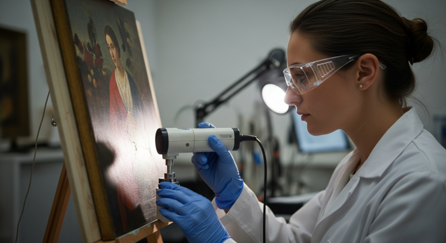
In Situ Analysis of Pigments on Artwork
One of the most significant advantages of raman spectroscopy is its ability to perform in situ analysis, meaning that the pigments can be analyzed directly on the artwork without the need for sampling. This is particularly important for fragile or valuable objects where any form of sampling is undesirable.
In situ analysis is typically performed using portable Raman spectrometers, which can be brought directly to the artwork. The instrument is focused on the area of interest, and the spectrum is acquired without any physical contact with the surface.
This non-destructive approach allows conservators to gain valuable information about the pigments used in the artwork without causing any damage. It also enables the analysis of multiple areas on the same artwork, providing a comprehensive understanding of the materials used by the artist.
However, in situ analysis can be challenging due to factors such as surface roughness, the presence of varnishes or coatings, and the potential for fluorescence interference. Careful selection of the laser wavelength and optimization of the instrument parameters are crucial for obtaining high-quality spectra in these situations.
Portable Raman spectrometers are designed to be lightweight and easy to use in the field. They often have integrated software for data acquisition and analysis.
When performing in situ analysis, it is important to carefully position the instrument to ensure that the laser beam is focused on the area of interest. A microscope objective is typically used for this purpose.
The presence of a varnish layer can significantly attenuate the Raman signal. In some cases, it may be necessary to use a higher laser power to obtain a detectable spectrum.
Fluorescence interference can be minimized by selecting a laser wavelength that does not excite the fluorescent molecules. Near-infrared (NIR) lasers are often used for this purpose.
Even with careful optimization, the spectra obtained from in situ analysis may be of lower quality than those obtained from prepared samples. However, the non-destructive nature of the technique makes it an invaluable tool for art conservation.
Advantages and Limitations of Raman Spectroscopy
Raman spectroscopy offers several advantages for the analysis of historical pigments. Its non-destructive nature is a major benefit, as it allows for the analysis of valuable and fragile artworks without causing damage.
The ability to perform in situ analysis is another significant advantage, as it eliminates the need for sampling and allows for the analysis of multiple areas on the same artwork. However, raman spectroscopy also has some limitations that must be considered.
- Fluorescence interference can obscure the Raman signal, making it difficult to obtain a clear spectrum.
- Some pigments, particularly those containing organic materials, may be susceptible to laser-induced degradation.
- The sensitivity of Raman spectroscopy can be lower compared to other analytical techniques, such as X-ray fluorescence (XRF).
- The interpretation of complex spectra can be challenging, especially when dealing with mixtures of pigments or the presence of degradation products.
- Reference databases may not always contain spectra for all possible pigments or variations thereof.
Despite its limitations, Raman spectroscopy remains a powerful tool for art conservation. Its advantages often outweigh the disadvantages, particularly when non-destructive analysis is required.
Fluorescence interference can sometimes be reduced by using techniques such as shifted excitation Raman difference spectroscopy (SERDS). This technique involves acquiring two Raman spectra with slightly different excitation wavelengths and subtracting them to remove the fluorescence background.
Laser-induced degradation can be minimized by using a lower laser power or by rastering the laser beam across the sample surface. This reduces the amount of energy deposited on any one spot.
The sensitivity of Raman spectroscopy can be improved by using a longer acquisition time or by averaging multiple spectra. However, this may not be practical for in situ analysis, where time is often limited.
The interpretation of complex spectra can be facilitated by using spectral deconvolution techniques. These techniques involve separating the overlapping peaks in the spectrum to identify the individual components.
Applications of Raman Spectroscopy in Art Conservation
Raman spectroscopy has a wide range of applications in the field of art conservation. It is used to identify the pigments used in paintings, sculptures, and other works of art, providing valuable information about the artist’s materials and techniques.
This information can be used to authenticate artworks, determine their provenance, and assess their condition. Raman spectroscopy can also be used to study the degradation of pigments over time, helping conservators understand the mechanisms of deterioration and develop appropriate conservation strategies.
For example, raman spectroscopy has been used to identify the pigments used in ancient Egyptian artifacts, revealing the use of materials such as Egyptian blue and ochre. It has also been used to study the fading of pigments in historical textiles, providing insights into the effects of light and humidity on these materials.
Furthermore, raman spectroscopy can be used to monitor the effectiveness of conservation treatments, such as cleaning and consolidation. By analyzing the pigments before and after treatment, conservators can assess the impact of the treatment on the materials and make adjustments as needed.
Raman spectroscopy aids in understanding the evolution of artistic practices. By identifying the pigments used over different periods, it’s possible to trace the development of painting techniques.
The technique can also be used to map the distribution of pigments within a painting. This provides insights into the artist’s working methods and the layering of different colors.
Raman spectroscopy is valuable in assessing the stability of pigments under different environmental conditions. This helps conservators to develop appropriate storage and display strategies.
The identification of degradation products can provide clues about the history of an artwork. It can reveal past conservation treatments or environmental exposures.
Raman spectroscopy is increasingly being used in conjunction with other analytical techniques, such as XRF and infrared spectroscopy. This multi-technique approach provides a more comprehensive understanding of the materials used in artworks.
Raman Spectroscopy for Analyzing Binding Media
While raman spectroscopy is most commonly associated with pigment analysis, it can also be used to identify the binding media used in artworks. Binding media are the substances that hold the pigment particles together and adhere them to the support, such as oil, egg tempera, or glue.
The identification of binding media can provide valuable information about the artist’s technique and the materials used in the artwork. Raman spectroscopy can be used to identify the characteristic vibrational modes of different binding media, allowing for their differentiation.
However, the analysis of binding media can be more challenging than pigment analysis due to the complexity of their chemical composition and the potential for degradation over time. The Raman spectra of binding media can be broad and overlapping, making it difficult to identify specific components.
In some cases, it may be necessary to use complementary analytical techniques, such as gas chromatography-mass spectrometry (GC-MS), to obtain a more complete understanding of the binding media composition. Despite these challenges, raman spectroscopy remains a valuable tool for the analysis of binding media in art conservation.
The Raman spectra of different binding media are often characterized by broad bands due to the amorphous nature of these materials. These bands can be difficult to interpret without reference spectra.
The degradation of binding media can lead to changes in their Raman spectra. Hydrolysis and oxidation can alter the chemical composition of the media, resulting in new peaks or shifts in existing peaks.
The presence of pigments can also interfere with the analysis of binding media. The Raman signals from the pigments can overlap with those from the media, making it difficult to distinguish between the two.
Despite these challenges, Raman spectroscopy can provide valuable information about the type of binding media used in an artwork. By comparing the spectrum of the sample to reference spectra of known binding media, it is often possible to identify the material with a reasonable degree of certainty.
The combination of Raman spectroscopy with other analytical techniques, such as GC-MS and Fourier transform infrared spectroscopy (FTIR), can provide a more complete picture of the composition of the binding media. This multi-technique approach is often necessary for the accurate identification of these complex materials.
Using Raman Spectroscopy to Detect Forgeries
Raman spectroscopy can play a crucial role in the detection of art forgeries. By identifying the pigments used in a work of art, it is possible to determine whether they are consistent with the purported date and origin of the piece.
If pigments are found that were not available during the time period the artwork is claimed to be from, it can raise serious doubts about its authenticity. For example, the presence of synthetic pigments that were not invented until the 19th or 20th centuries in a painting supposedly created in the 17th century would be a clear indication of forgery.
Raman spectroscopy can also be used to detect the use of modern materials in restoration or retouching, which may not be immediately apparent to the naked eye. This can help to uncover fraudulent attempts to enhance the value of an artwork by concealing damage or alterations.
However, it is important to note that pigment analysis alone is not always sufficient to definitively prove or disprove the authenticity of an artwork. Other factors, such as the style of the work, the provenance, and the results of other analytical techniques, must also be considered.
The identification of anachronistic pigments is a strong indicator of forgery. However, it is important to rule out the possibility of later additions or restorations before drawing conclusions.
Raman spectroscopy can also be used to detect the presence of modern binding media or varnishes. These materials may not have been available during the purported time period of the artwork.
The absence of expected pigments can also be a sign of forgery. If an artwork is claimed to be from a period when certain pigments were commonly used, their absence may raise suspicions.
The use of Raman spectroscopy in conjunction with other analytical techniques, such as X-ray diffraction (XRD) and scanning electron microscopy (SEM), can provide a more comprehensive assessment of the authenticity of an artwork.
Ultimately, the determination of authenticity is a complex process that requires careful consideration of all available evidence. Raman spectroscopy is a valuable tool in this process, but it should not be used in isolation.
Case Studies: Raman Spectroscopy in Action
Numerous case studies demonstrate the effectiveness of raman spectroscopy in art conservation. One notable example is the analysis of the Vinland Map, a controversial map purported to be a pre-Columbian depiction of North America.
Raman spectroscopy revealed the presence of titanium dioxide in the ink, a pigment that was not widely available until the 20th century. This finding cast serious doubt on the map’s authenticity, suggesting that it was a modern forgery.
Another case study involves the analysis of a collection of ancient Roman frescoes. Raman spectroscopy was used to identify the pigments used in the frescoes, providing insights into the materials and techniques employed by Roman artists.
The analysis revealed the use of pigments such as المصري blue, ochre, and vermilion, which were commonly used in Roman art. This information helped to confirm the authenticity of the frescoes and provided valuable information for their conservation.
In the study of medieval illuminated manuscripts, Raman spectroscopy helped identify the pigments used in the intricate illustrations. This allowed art historians to understand the trade routes and artistic influences of the time.
Raman analysis of Vincent van Gogh’s paintings has revealed the presence of light-sensitive pigments that have degraded over time. This information is crucial for developing appropriate conservation strategies to prevent further damage.
The examination of ancient Chinese ceramics using Raman spectroscopy has identified the different glazes and pigments used in their production. This provides insights into the technological advancements of the time.
Raman spectroscopy has been used to analyze the pigments in the Dead Sea Scrolls, helping to understand the materials used in their creation and to assess their state of preservation.
These case studies demonstrate the diverse applications of Raman spectroscopy in art conservation and its ability to provide valuable information about the materials, techniques, and history of artworks.
Conclusion
Raman spectroscopy is a powerful and versatile tool for the analysis of historical pigments and other materials in art conservation. Its non-destructive nature, ability to perform in situ analysis, and capacity to provide detailed information about the chemical composition of materials make it an indispensable technique for conservators, art historians, and scientists.
By providing insights into the materials and techniques used in artworks, raman spectroscopy helps to inform conservation strategies, authenticate artworks, and deepen our understanding of the artistic heritage. As technology advances, raman spectroscopy will likely play an even greater role in the field of art conservation, helping to preserve and protect our cultural treasures for future generations.
The continued development of portable Raman spectrometers will further enhance the accessibility of this technique. This will enable more widespread in situ analysis of artworks in museums and archaeological sites.
Advances in data processing and spectral analysis techniques will improve the accuracy and reliability of Raman spectroscopy. This will allow for the identification of even trace amounts of pigments and other materials.
The integration of Raman spectroscopy with other analytical techniques will provide a more comprehensive understanding of the materials used in artworks. This multi-technique approach will become increasingly common in art conservation research.
Raman spectroscopy will continue to play a vital role in preserving our cultural heritage. By providing insights into the materials and techniques used in artworks, it helps us to understand and appreciate the artistic achievements of the past.